
Fig. 2
- ID
- ZDB-FIG-160517-45
- Publication
- Tian et al., 2016 - Extraocular Source of Oligodendrocytes Contribute to Retinal Myelination and Optokinetic Responses in Zebrafish
- Other Figures
- All Figure Page
- Back to All Figure Page
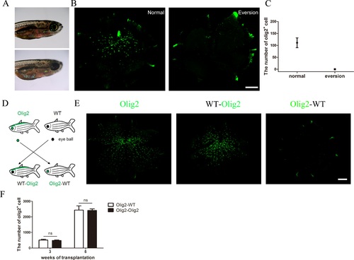
The origin of OLs in zebrafish retinas. (A) An illustrative picture shows the normal and eye eversion zebrafish. (B, C) Whole mount and quantification of the number of OLs in normal olig2 zebrafish retinas and the retinas after eye eversion 2 weeks later. (D) An illustrative picture shows the exchange of eyeballs in WT and olig2 zebrafish. (E) Whole-mount retinas show OLs in normal olig2 zebrafish eyes, olig2 zebrafish with WT eyes, and WT zebrafish with olig2 eyes at 3 weeks after eye eversion. (F) Quantification of the number of OLs in normal olig2 zebrafish eyes and olig2 zebrafish with WT eyes (n = 5 and n = 7, respectively) at 3 and 5 weeks after eye eversion. Scale bars: (B) = 150 µm, (E) = 200 µm. |