
Fig. 1
- ID
- ZDB-FIG-160517-44
- Publication
- Tian et al., 2016 - Extraocular Source of Oligodendrocytes Contribute to Retinal Myelination and Optokinetic Responses in Zebrafish
- Other Figures
- All Figure Page
- Back to All Figure Page
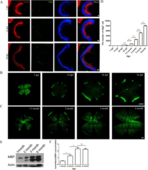
Development of myelination in zebrafish retinas. (A) Cryosections of 7- to 28-dpf zebrafish retinas were stained with MBP and a tubulin antibody. Myelin basic protein appeared at 28 dpf. (B, C) Whole-mount olig2 zebrafish retinas at the age of 20 to 36 dpf and 1.5 to 6.0 months. The olig2+ cells, outside the nerve fiber layer, were not OLs at 7 and 14 dpf. (D) Quantification of the number of OLs (n = 10, 10, 44, 21, 14, 13, 10, and 8, respectively) in zebrafish retinas from 7 dpf to 6 months. (E, F) Myelin basic protein Western blot of zebrafish retinas and its quantitative analysis at age 1.5 to 6.0 months. Scale bars: (A) = 100 µm; (B, C) = 200 µm. |