
Fig. 6
- ID
- ZDB-FIG-160517-49
- Publication
- Tian et al., 2016 - Extraocular Source of Oligodendrocytes Contribute to Retinal Myelination and Optokinetic Responses in Zebrafish
- Other Figures
- All Figure Page
- Back to All Figure Page
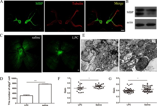
The OLs in zebrafish retinas are functional. (A) Cryosections of normal (right eye) and demyelinated (left eye) zebrafish retinas were immunolabeled with MBP and a tubulin antibody. (B) Myelin basic protein Western blot of normal and demyelinated zebrafish retinas. (C) Whole mount of normal and demyelinated olig2 zebrafish retinas. (D) Quantification of the number of OLs in the normal and demyelinated olig2 zebrafish retinas. (E) Electron micrographs of RGC axons in normal and LPC-induced demyelinated zebrafish retinas. (F, G) Optokinetic reflex behavior test of saline-treated and LPC-induced demyelinated zebrafish at the speed of 12 (n = 19, 18) and 24 rpm (n = 38, 41). Scale bars: (A) = 100 µm, (C) = 200 µm, and (E) = 200 nm. |