- Title
-
Crispld2 is required for neural crest cell migration and cell viability during zebrafish craniofacial development
- Authors
- Swindell, E.C., Yuan, Q., Maili, L.E., Tandon, B., Wagner, D.S., Hecht, J.T.
- Source
- Full text @ Genesis
|
NCC migration is disorganized in MO3 morphants. Time-lapse live cell imaging captures of sox10:GFP embryos showing migrating NCC cells at 16 (a, b), 22 (c, d), and 28 hpf (e, f). UIC (a, c, and e) and MO3-injected embryos (b, d, and f) showing a dorsal view of migrating NCCs. White arrows point to abnormally migrating NCCs. (g) Quantification of number of cells crossing the midline in UIC, MO3 and control MM-MO3 injected embryos. EXPRESSION / LABELING:
PHENOTYPE:
|
|
Aberrant oral cartilage in MO3 morphants. Time-lapse live cell imaging captures of sox10:GFP embryos showing migrating NCC cells at 48 (a, b), 60 (c, d), and 72 hpf (e, f). UIC (a, c, and e) and MO3-injected embryos (b, d, and f) showing a ventral view of migrating NCCs. White arrows point to loss of normal migration and abnormal formation of presumptive cartilage elements. EXPRESSION / LABELING:
PHENOTYPE:
|
|
Increased apoptosis is present in the head of MO3 injected embryos. Co-injection of MO3 with p53MO and whole mount TUNEL staining shows partial inhibition of MO3-induced apoptosis. Lateral view of UIC (a–f), MO3-injected embryos (g–l), MO3/p53MO co-injected embryos (m–r), and control MM-MO3-injected embryos (s–x), at 16 hpf (a, g, m, s), 24 hpf (b, h, n, t), 36 hpf (c, i, o, u), 48 hpf (d, j, p, v), 60 hpf (e, k, q, w), and 72 hpf (f, l, r, x). PHENOTYPE:
|
|
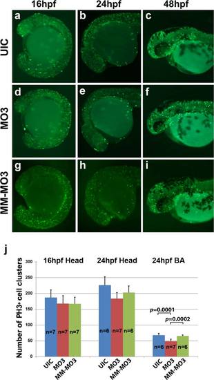
Cell proliferation is unchanged in MO3 injected embryos. Whole mount immunohistochemistry with mitotic marker anti-phosphohistone H3 shows proliferating cells in developing embryos. UIC (a–c), MO3 (d–f), and control MM-MO3-injected embryos (g–i) embryos at 16 hpf (a, d, g), 24 hpf (b, e, h), and 48 hpf (c, f, i). (j) Quantification of number of proliferating cells in UIC, MO3, and control MM-MO3 injected embryos at 16 and 24 hpf. PHENOTYPE:
|
|
Crispld2 is highly expressed in zebrafish oropharynx at 48 hpf. (a) Schematic presentation of the location and orientation of transverse sections from rostral to caudal. (b) Top panels show Crispld2 immunostaining through zebrafish oropharynx (bright green) from rostral to caudal at 20× magnification. (c) Enlarged images of oropharynx area (rectangle) of embryos shown in B. EXPRESSION / LABELING:
|
|
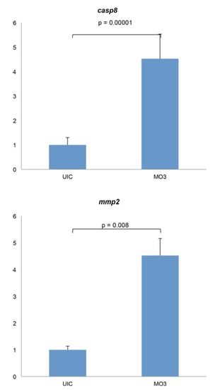
Cell death related gene expression is up-regulated in Crispld2 knockdown embryos. Both mmp2 and casp8 show marked increase in mRNA expression levels in knockdown embryos at 16 hpf. P-value less than 0.05 denotes statistical significance. EXPRESSION / LABELING:
PHENOTYPE:
|


