
- Title
-
Cyclosporin A Disrupts Notch Signaling and Vascular Lumen Maintenance
- Authors
- Pandey, R., Botros, M.A., Nacev, B.A., Albig, A.R.
- Source
- Full text @ PLoS One
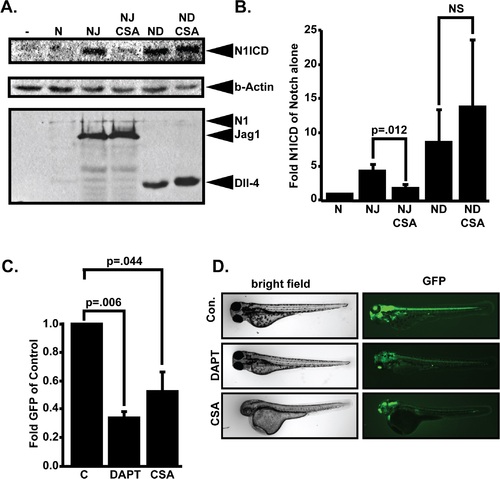
CSA blocks Notch signaling. (A) Effect of CSA on Notch signaling in vitro. 293T cells were transfected with various combinations of myc-tagged murine Notch1 (N), JAG1 (J), or Delta-like 4 (D) and treated with either 0.1% DMSO or 10µM CSA. Whole cell lysates were fractionated through SDS-PAGE gels and western blotted with anti-Val1744 antibody to detect cleaved Notch1 NICD fragments (N1ICD). Stripped blots were re-blotted with β-actin or 9E10 anti-myc antibodies to control for protein loading and expression of various transfected cDNAs. Shown are representative western blots from a single experiment that was performed five times in its entirety. (B) Western blot quantitation comparing N1ICD levels in cells transfected with Notch1 alone to cells transfected with combinations of Notch and JAG1 or Dll4 in the presence or absence of CSA. Displayed data represent the mean +/- SE of five individual experiments. P-values were calculated with the Student’s t-test. (C) Effects of CSA on Notch activity in vivo. Tp1bglob:eGFP embryos which express GFP from a tandem array of 12 Notch responsive RBP-Jk binding sites were incubated in either 0.1% DMSO, 10µM DAPT, or 10µM CSA. 48 hours later, GFP signal intensity was quantified in whole, live embryos. Data shown represents the mean +/- SE of 4 individual experiments. P-values were determined by student’s t-test. (D) Representative pictures of Tp1bglob:eGFP zebrafish embryos incubated with 10M DAPT or 10M CSA and imaged by fluorescent microscopy. |
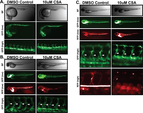
Cyclosporin-A destabilizes vascular lumen structures in zebrafish embryos. Freshly laid Fli1-GFP/GATA1-RFP zebrafish embryos were incubated in 10µM CSA or DMSO vehicle control for one, two, or four days. Whole embryo brightfield imaging was used to monitored gross morphology. Development of the vascular system was monitored by fluorescent microscopy of endothelial specific GFP expression. Circulatory flow was monitored by fluorescent microscopy of red-blood cell specific RFP expression. (A) Effects of CSA on 1dpf embryos. 1 day after CSA treatment, brightfield imaging of zebrafish embryos (top panel) was unable to distinguish any significant developmental impact of CSA on gross embryo morphology. Microangiogram analysis revealed similar development of the primitive vascular system including sprouting intersegmental vessels. (B) Effects of CSA on 2 dpf embryos. Zebrafish embryos treated with CSA for two days displayed no obvious signs of developmental abnormality in bright field images. Low power GFP imaging revealed an apparently normal vascular system, however RFP imaging revealed a distinct lack of blood flow throughout the embryo. High power GFP imaging revealed a lack of vascular lumen structures in ISV structures (arrows). (C) Effect of CSA on 4 dpf embryos. After four days of CSA treatment, no vascular luminal structures (arrows) or blood flow was evident in CSA treated embryos. |

Inhibition of cyclophilin A or calcineurin/NFAT destabilizes lumen structure. (A) Freshly laid Fli1-GFP / GATA1-RFP embryos were incubated in 0.1% DMSO, 2µM FK506, or 40µM N-MeVal-4-CsA (CSA-Analog) for two days. Fluorescent imaging was used to monitor overall vascular development (GFP) and blood flow (RFP). Similar to CSA, neither FK506 nor N-MeVal-4-CSA had an impact on gross morphology or overall vascular development however both drugs blocked blood flow. |

Notch inhibition partially rescues CSA induced vascular malfunction. (A) Effect of CSA and DAPT on vascular function in zebrafish embryos. Freshly laid Fli1-GFP / GATA1-RFP zebrafish embryos were incubated in 0.1% DMSO (Control), 10µM CSA, 15µM DAPT, or 10µM CSA + 15µM DAPT for 2 days. Bright field imaging revealed no gross morphological abnormalities in either CSA or DAPT treated fish, however CSA + DAPT treated fish experienced an acute curvature. GFP imaging revealed a lack of lumen structures in the ISV (white arrowheads) and aortic vessels (red arrowheads) of CSA treated fish. DAPT treated fish displayed normal luminal structure and blood flow. CSA + DAPT treated embryos had luminal structures (arrowheads) and blood flow similar to control or DAPT alone treated embryos. Shown are representative results from a single experiment that was performed five times in its entirety. (B) Quantitative analysis of blood flow in zebrafish treated with CSA, DAPT, or CSA + DAPT. Data shown represent the average +/- SE of five individual experiments. P-values were determined by student’s t-test. |