
Fig. 3
- ID
- ZDB-FIG-150504-9
- Publication
- Pandey et al., 2015 - Cyclosporin A Disrupts Notch Signaling and Vascular Lumen Maintenance
- Other Figures
- All Figure Page
- Back to All Figure Page
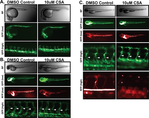
Cyclosporin-A destabilizes vascular lumen structures in zebrafish embryos. Freshly laid Fli1-GFP/GATA1-RFP zebrafish embryos were incubated in 10µM CSA or DMSO vehicle control for one, two, or four days. Whole embryo brightfield imaging was used to monitored gross morphology. Development of the vascular system was monitored by fluorescent microscopy of endothelial specific GFP expression. Circulatory flow was monitored by fluorescent microscopy of red-blood cell specific RFP expression. (A) Effects of CSA on 1dpf embryos. 1 day after CSA treatment, brightfield imaging of zebrafish embryos (top panel) was unable to distinguish any significant developmental impact of CSA on gross embryo morphology. Microangiogram analysis revealed similar development of the primitive vascular system including sprouting intersegmental vessels. (B) Effects of CSA on 2 dpf embryos. Zebrafish embryos treated with CSA for two days displayed no obvious signs of developmental abnormality in bright field images. Low power GFP imaging revealed an apparently normal vascular system, however RFP imaging revealed a distinct lack of blood flow throughout the embryo. High power GFP imaging revealed a lack of vascular lumen structures in ISV structures (arrows). (C) Effect of CSA on 4 dpf embryos. After four days of CSA treatment, no vascular luminal structures (arrows) or blood flow was evident in CSA treated embryos. |