
|
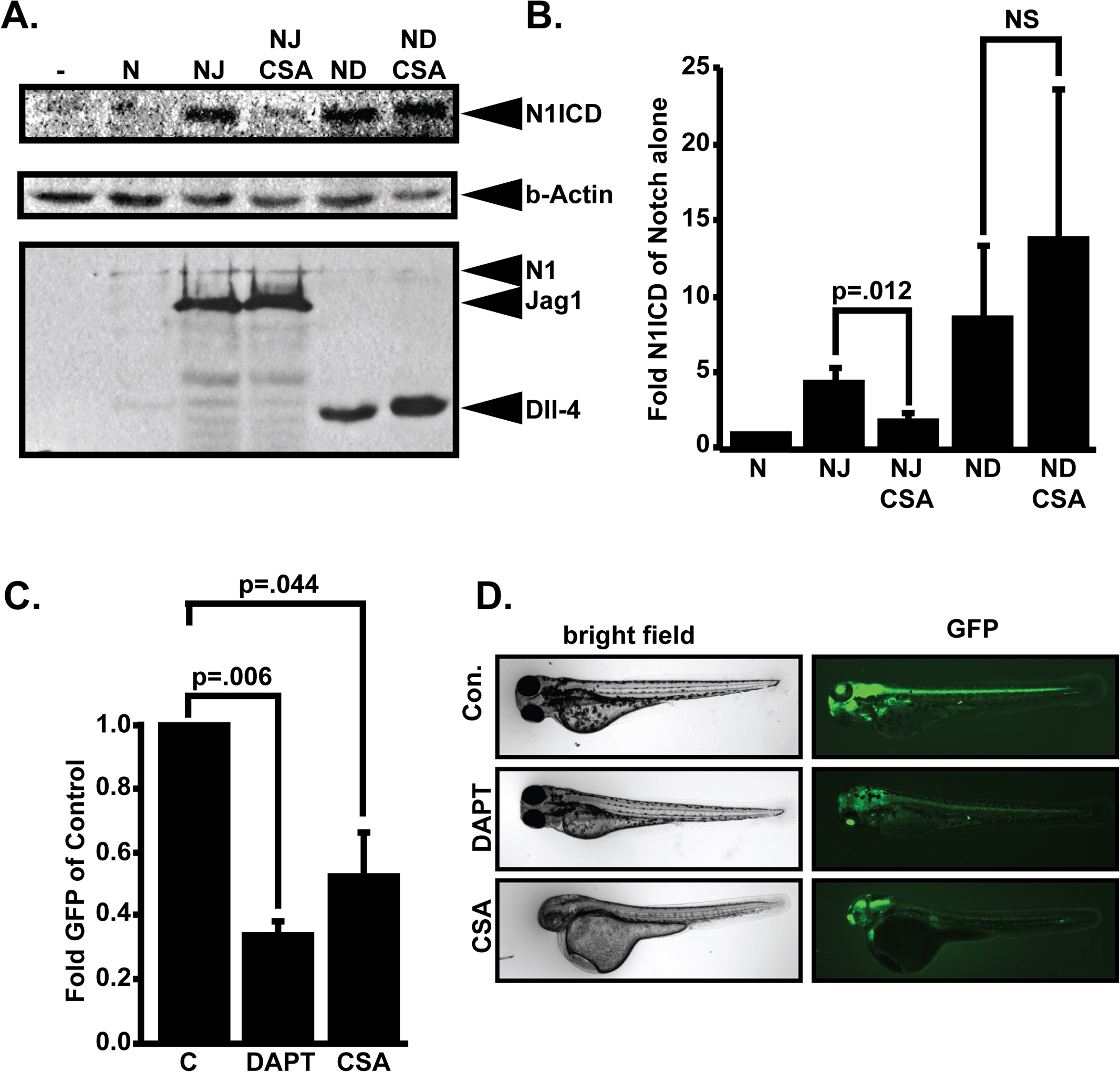
Fig. 1 CSA blocks Notch signaling.
(A) Effect of CSA on Notch signaling in vitro. 293T cells were transfected with various combinations of myc-tagged murine Notch1 (N), JAG1 (J), or Delta-like 4 (D) and treated with either 0.1% DMSO or 10µM CSA. Whole cell lysates were fractionated through SDS-PAGE gels and western blotted with anti-Val1744 antibody to detect cleaved Notch1 NICD fragments (N1ICD). Stripped blots were re-blotted with β-actin or 9E10 anti-myc antibodies to control for protein loading and expression of various transfected cDNAs. Shown are representative western blots from a single experiment that was performed five times in its entirety. (B) Western blot quantitation comparing N1ICD levels in cells transfected with Notch1 alone to cells transfected with combinations of Notch and JAG1 or Dll4 in the presence or absence of CSA. Displayed data represent the mean +/- SE of five individual experiments. P-values were calculated with the Student’s t-test. (C) Effects of CSA on Notch activity in vivo. Tp1bglob:eGFP embryos which express GFP from a tandem array of 12 Notch responsive RBP-Jk binding sites were incubated in either 0.1% DMSO, 10µM DAPT, or 10µM CSA. 48 hours later, GFP signal intensity was quantified in whole, live embryos. Data shown represents the mean +/- SE of 4 individual experiments. P-values were determined by student’s t-test. (D) Representative pictures of Tp1bglob:eGFP zebrafish embryos incubated with 10M DAPT or 10M CSA and imaged by fluorescent microscopy.