
|
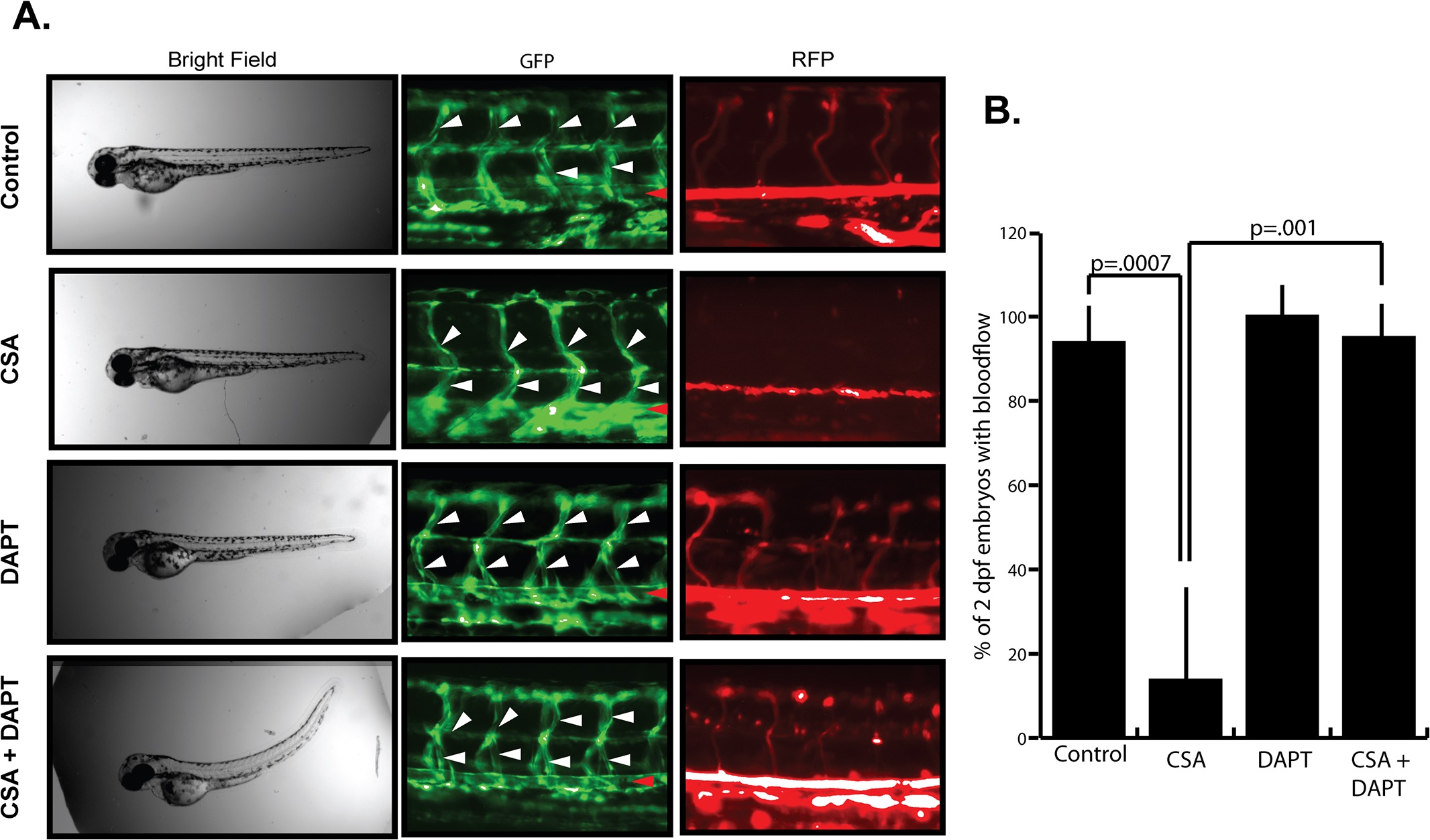
Fig. 5 Notch inhibition partially rescues CSA induced vascular malfunction.
(A) Effect of CSA and DAPT on vascular function in zebrafish embryos. Freshly laid Fli1-GFP / GATA1-RFP zebrafish embryos were incubated in 0.1% DMSO (Control), 10µM CSA, 15µM DAPT, or 10µM CSA + 15µM DAPT for 2 days. Bright field imaging revealed no gross morphological abnormalities in either CSA or DAPT treated fish, however CSA + DAPT treated fish experienced an acute curvature. GFP imaging revealed a lack of lumen structures in the ISV (white arrowheads) and aortic vessels (red arrowheads) of CSA treated fish. DAPT treated fish displayed normal luminal structure and blood flow. CSA + DAPT treated embryos had luminal structures (arrowheads) and blood flow similar to control or DAPT alone treated embryos. Shown are representative results from a single experiment that was performed five times in its entirety. (B) Quantitative analysis of blood flow in zebrafish treated with CSA, DAPT, or CSA + DAPT. Data shown represent the average +/- SE of five individual experiments. P-values were determined by student’s t-test.