- Title
-
The epigenetic factor Kmt2a/Mll1 regulates neural progenitor proliferation and neuronal and glial differentiation
- Authors
- Huang, Y.C., Shih, H.Y., Lin, S.J., Chiu, C.C., Ma, T.L., Yeh, T.H., Cheng, Y.C.
- Source
- Full text @ Dev. Neurobiol.
|
kmt2a is expressed in the developing nervous system and the expression is required for neural development. (A) kmt2a expression was detected by in situ hybridization in the developing nervous system during zebrafish embryogenesis. The embryo stages are shown in the bottom left corner of each panel. All panels are lateral view, with anterior to the left except 0.75 hpf, 5.3 hpf, and 8 hpf. Ubiquitous expression of kmt2a was detected from 0.75 hpf to 16 hpf. Strong expression was detected in the entire nervous system at 24 hpf and later became restricted to specific brain areas, persisting until the final stage that was analyzed (48 hpf). di, diencephalon; fb, forebrain; hb, hindbrain; mb, midbrain; sp, spinal cord; t, tectum; hpf, hours postfertilization. (B) Interference with Kmt2a expression using kmt2a morpholino or kmt2aN84 resulted in brain malformation (arrowheads). Note, however, that the midbrain-hindbrain boundaries (white arrowheads) and somitic boundaries are unaffected. Scale bars: 200 µm. EXPRESSION / LABELING:
PHENOTYPE:
|
|
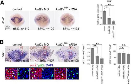
Defective Kmt2a expression decreased the proliferation of neural progenitors. (A) In situ hybridization of 75%-epiboly embryos showed that sox2 expression was downregulated by the kmt2a morpholino or kmt2aN84 cRNA, as confirmed by qPCR analysis on the right. (B) Embryos were flat-mounted and double-labeled with sox2 and phospho-histone H3 antibody. The bottom panels are representative of the enlargement regions of the upper panels, as indicated. sox2-expressing cells were pseudo-colored with fluorescent red and counterstained with phospho-histone H3 antibody for immunohistochemistry (fluorescent green) to locate proliferating neural progenitor cells (arrowheads). Injection of the kmt2a morpholino or kmt2aN84 decreased proliferation of neural progenitors. The proportions of phospho-histone H3- and sox2-positive cells among the total sox2-positive cells were quantified, as shown on the right. Note that in the Kmt2a-deficient embryos, no significant deviation was observed in the proportions of phosphohistone H3-positive and sox2-negative cells in the sox2-negative cells counted in adjacent surface ectoderm. EXPRESSION / LABELING:
PHENOTYPE:
|
|
kmt2a morpholino or kmt2aN84 cRNA injection had no effect on neural progenitor apoptosis. Apoptotic neural progenitor cells were labeled for sox2 (fluorescent red) and activated caspase-3 antibody (fluorescent green). As shown in E, apoptotic neural progenitor cells were quantified by counting the proportions of activated caspase-3- and sox2-positive (or -negative) cells among the total sox2-positive (or -negative) cells. *p < 0.05; **p < 0.01; n.s., not significant. Scale bar: 100 µm, applies to all panels. EXPRESSION / LABELING:
|
|
Disrupted Kmt2a expression causes aberrant formation of neuronal precursors and mature neurons. (A) At the bud stage, the expression level of neurogenin1 was significantly decreased in embryos injected with the kmt2a morpholino or kmt2aN84 cRNA injected embryos in comparison to the controls. (B) qPCR analysis confirmed the results obtained by in situ hybridization in A. (C and E) The upper panels are enlargements of the hindbrain region, anterior to the top; and the bottom panels present lateral views of the enlargements of the 3–9-somite levels of the spinal cord. (C) HuC/D-expressing post-mitotic neurons increased massively in kmt2a morpholino or kmt2aN84 cRNA injected embryos, as shown by immunohistochemical analysis with anti-HuC/D antibody at 24 hpf. Note the ectopic HuC/D expression in the ventricular zone (arrowheads). (D) Levels of HuC/D expression were confirmed by Western blot analysis and were quantified. (E) At 24 hpf, neurogenin1 expression was reduced significantly by the kmt2a morpholino and kmt2aN84. (F) qPCR analysis further confirmed the decreased expression of neurogenein1 at 24 hpf in E. *p < 0.05; **p < 0.01. |
|
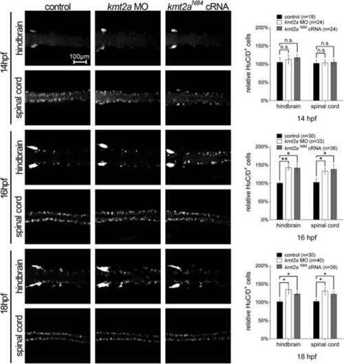
Kmt2a-deficient embryos exhibit upregulated HuC/D expression. Significant upregulation of HuC/D signals, as analyzed by immunohistochemistry using an anti-HuC/D antibody, can be detected at 16 hpf and 18 hpf in embryos injected with either the kmt2a morpholino or kmt2aN84 cRNA. Quantitative data are presented as the mean ± standard deviation normalized to the number of controls. *p < 0.05; **p < 0.01; n.s., not significant. Scale bar: 100 µm, applies to all panels. EXPRESSION / LABELING:
PHENOTYPE:
|
|
Disrupted Kmt2a expression causes decreased glial precursors and derivatives. The expression of slc1a3a, oilg1, and mag were analyzed by in situ hybridization (A), and Gfap was examined in Tg(gfap:egfp) transgenic embryos (C). Injection of the kmt2a morpholino or kmt2aN84 cRNA downregulated the expression of all these glial markers. (B) qPCR analysis confirmed the results obtained by in situ hybridization in A. (D) Levels of GFP expression in C were confirmed by Western blot analysis and were quantified. Quantitative data are presented as the means ± standard deviations. *p < 0.05; **p < 0.01; n.s., not significant. EXPRESSION / LABELING:
PHENOTYPE:
|


