
- Title
-
Slitrk gene duplication and expression in the developing zebrafish nervous system
- Authors
- Round, J., Ross, B., Angel, M., Shields, K., and Lom, B.
- Source
- Full text @ Dev. Dyn.

Seven slitrk genes are expressed in developing and adult zebrafish. Nonquantitative reverse transcriptase-polymerase chain reaction (RT-PCR) was performed on total RNA isolated from whole zebrafish ranging in age from 12 hr postfertilization (hpf) to adulthood (90+ days postfertilization [dpf]). Transcripts corresponding to seven of eight predicted slitrks (1, 2, 3a, 3b, 4, 5a, 6) were detected from 48 dpf to adulthood, with slitrk6 detected as early as 24 hpf. Transcripts corresponding to slitrk5b were not detected at any age examined. To control for RNA integrity, β-actin was detected at all time-points examined (βactin+RT) and the absence of genomic DNA was confirmed by RT-PCR for β-actin in the absence of reverse transcriptase (βactin-RT). |
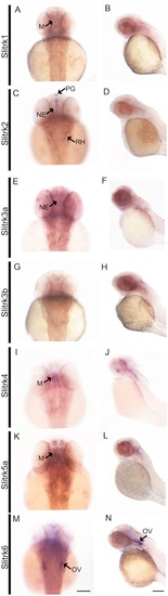
A-N Slitrk expression at 48 hr postfertilization (hpf) is observed in the developing nervous system. In situ hybridization performed on whole-mount zebrafish embryos at 48 hpf demonstrates differential expression patterns throughout the developing brain of seven different slitrks. A dorsal view of the head (left) and lateral view of the whole embryo (right) are provided for each slitrk gene. M, midbrain; NE, neuroepithelium; OV, otic vesicle; PG, pineal gland; RH, rhombomeres of the hindbrain. Scale bars, 50 μm. EXPRESSION / LABELING:
|
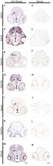
A-N Slitrk expression at 72 hr post fertilization is observed in the developing retina and midbrain. In situ hybridization at 72 hpf zebrafish highlights differential expression patterns of specific slitrks in sections through retina and midbrain treated with antisense slitrk probes (A,C,E,G,I,K,M). Most slitrks are expressed in the hypothalamus and tegmentum (A,C,G,I,M), and slitrk2 and slitrk3b are detected in the neuroepithelium (C,G). Lack of staining with corresponding sense probes (B,D,F,H,J,L,N) indicates antisense probe specificity. Ha, habenula; Hy, hypothalamus; NE, neuroepithelium; OT, optic tectum; T, tegmentum. Scale bars, 50 μm. EXPRESSION / LABELING:
|
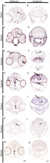
A-N Slitrk expression at 10 days post fertilization is observed in the developing midbrain and hindbrain. In situ hybridization of 10 dpf zebrafish sections demonstrates differential expression patterns of the seven expressed slitrks in the midbrain (A,C,E,G,I,K,M) and hindbrain (B,D,F,H,J,L,N). Multiple slitrks are expressed in the thalamus (A,C,G,I,K) and hypothalamus (G,I,M), and slitrk5b is detected in the optic tectum. Notably, slitrk3b is highly expressed in the cerebellar valvula and ventricular recess of the hypothalamus. CeP, cerebellar plate; Hy, hypothalamus; OT, optic tectum; T, tegmentum; Th, thalamus; VA, cerebellar valvula; VR, ventricular recess of hypothalamus. Scale bars, 50 μm. EXPRESSION / LABELING:
|
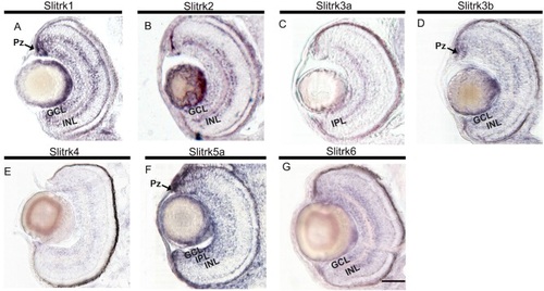
A-G Slitrk expression in the developing retina. In situ hybridization of sections of the zebrafish retina at 72 hpf demonstrates differential slitrk expression in multiple areas of the retina including ganglion cell layer (GCL), inner nuclear layer (INL), inner plexiform layer (IPL), and proliferative zone (Pz). Scale bars, 50 μm. EXPRESSION / LABELING:
|

A-G Slitrk expression in the developing spinal cord. In situ hybridization of 72 hpf zebrafish spinal cord sections reveals most but not all Slitrks are expressed throughout the developing spinal cord. SC, spinal cord. Scale bars, 50 μm. EXPRESSION / LABELING:
|

Unillustrated author statements EXPRESSION / LABELING:
|