|
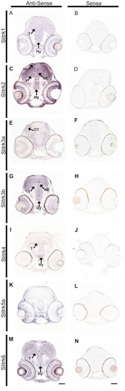
A-N Slitrk expression at 72 hr post fertilization is observed in the developing retina and midbrain. In situ hybridization at 72 hpf zebrafish highlights differential expression patterns of specific slitrks in sections through retina and midbrain treated with antisense slitrk probes (A,C,E,G,I,K,M). Most slitrks are expressed in the hypothalamus and tegmentum (A,C,G,I,M), and slitrk2 and slitrk3b are detected in the neuroepithelium (C,G). Lack of staining with corresponding sense probes (B,D,F,H,J,L,N) indicates antisense probe specificity. Ha, habenula; Hy, hypothalamus; NE, neuroepithelium; OT, optic tectum; T, tegmentum. Scale bars, 50 μm.
|