
- Title
-
Modulation by Cocaine of Dopamine Receptors through miRNA-133b in Zebrafish Embryos
- Authors
- Barreto-Valer, K., López-Bellido, R., Macho Sánchez-Simón, F., and Rodríguez, R.E.
- Source
- Full text @ PLoS One

ZFIN is incorporating published figure images and captions as part of an ongoing project. Figures from some publications have not yet been curated, or are not available for display because of copyright restrictions. EXPRESSION / LABELING:
PHENOTYPE:
|
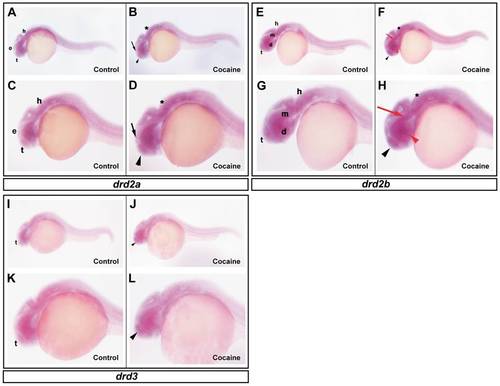
Effects of cocaine on the spatial distribution of drd2a, drd2b and drd3. Lateral view showing the expression of drd2a in embryos of 24 hpf. Control group (A and C) and cocaine group (B and D). drd2a is expressed in the epiphysis (e), tegmentum (t) and hindbrain (h) (A and C). Embryos exposed to cocaine (B and D) show an increase in the epiphysis (black arrow), tegmentum (black arrow head) and hindbrain (asterisk). Expression of drd2b in zebrafish embryos at 24 hpf. Control group (E and G) and cocaine group (F and H). drd2b is expressed in the tegmentum (t), diencephalon (d), mesencephalon (m) and in hindbrain (h) regions. Exposure to cocaine slightly increased the expression of drd2b in the regions mentioned (black arrow head, red arrow head, red arrow and asterisk, respectively). drd3 expression in zebrafish embryos at 24 hpf. Control group (I and K) and cocaine group (J and L). The expression of drd3 is seen mainly in tegmentum (t). Exposure to cocaine increased the expression of drd3 in tegmentum (black arrow head). Scale bars = 300 μm and 6X of magnification (A, B, E, F, I and J); 250 μm and 12X magnification (C, D, G, H, K and L). d: diencephalon (red arrow head), e: epiphysis (black arrow); h: hindbrain (asterisk); m: mesencephalon (red arrow); t: tegmentun (black arrowhead). |
|
ZFIN is incorporating published figure images and captions as part of an ongoing project. Figures from some publications have not yet been curated, or are not available for display because of copyright restrictions. |
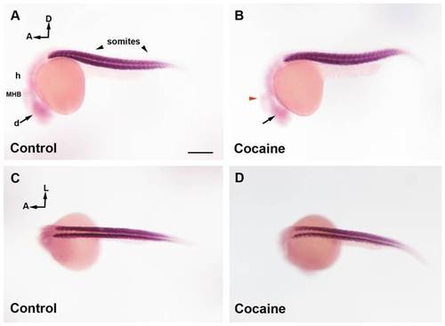
miR-133b distribution in zebrafish embryos at 24 hpf by whole-mount ISH. Control Group (A and C) and cocaine group (B and D). In a lateral view of miR-133b expression (A), the miRNA is mainly localized in somites and weakly in the brain (diencephalon, midbrain, MHB and hindbrain). Embryos exposed to cocaine (B) show a decrease in diencephalic expression (black arrow), and MHB (red arrowhead). A dorsal view of miR-133b (C) shows that this miRNA is mainly present in somites, although it is difficult to determine whether cocaine affects the expression of miR-133b in this area (D). Scale bars = 300 μm. A, B; C; D = 6X. A: anterior; D: dorsal; L: lateral; d: diencephalon; m: midbrain, h: hindbrain; MHB: Midbrain Hindbrain Boundary, sm: skeletal muscle. |
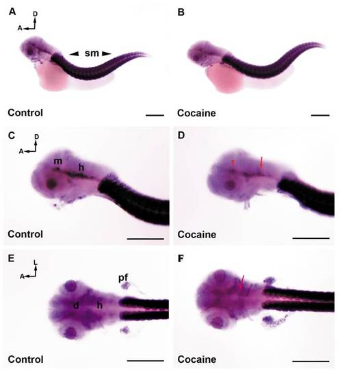
miR-133b distribution in zebrafish embryos at 48 hpf by whole-mount in situ hybridization (ISH). Control Group (A, C and E) and cocaine group (B, D and F). The expression of miR-133b is mostly found in the skeletal muscles (A, C and E) and to a lesser extent in the CNS (A, C and E). miR-133b is weakly expressed in the diencephalon, midbrain, and hindbrain. Embryos exposed to cocaine (B, D, and F) show a decrease mainly in the CNS (D), midbrain (red arrow head) and hindbrain (red arrow), while in the diencephalon (black arrow) the decrease less patent. The effect of cocaine on the expression of miR-133b in muscle is difficult to determine (D and F). Scale bars: 300 μm. A, B = 5X, and C, D, E, F = 10X. A: anterior; D: dorsal; L: lateral; d: diencephalon; m: midbrain; h: hindbrain; MHB: Midbrain Hindbrain Boundary; sm: skeletal muscle. |