
- Title
-
Cloning and expression of a zebrafish SCN1B ortholog and identification of a species-specific splice variant
- Authors
- Fein, A.J., Meadows, L.S., Chen, C., Slat, E.A., and Isom, L.L.
- Source
- Full text @ BMC Genomics

In situ hybridization. A – D: Fish stained at 24 hpf. A. Staining is apparent in the olfactory placode (OP) and the midbrain (Mb). B. Dorsal mount showing staining in the trigeminal neuron (Tg) and in the rhombomeres of the hindbrain (Hb). C. Staining in spinal cord and skeletal muscle (sm). D. Higher magnification of Rohon Beard cells (RB) flanking skeletal muscle (sm). E. Fish at 48 hpf with staining in the Rohan Beard cells of the spinal cord (SC) and in the skeletal muscle. F. Staining throughout the brain, at the olfactory pits (OP), in the layers of the retina, and in the trigeminal ganglion (Tg) of fish at 72 hpf. EXPRESSION / LABELING:
|
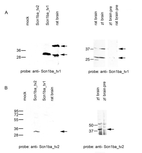
Antibody characterization. A. Left panel: Western blot probed with anti-Scn1ba_tv1. Lane 1: mock transfected Chinese hamster lung 1610 cells; Lane 2: Chinese hamster lung 1610 cells transiently transfected with scn1ba_tv2 cDNA; Lane 3: Chinese hamster lung 1610 cells transiently transfected with scn1ba_tv1 cDNA; Lane 4: 5 μg rat brain membranes. Arrows indicate immunoreactive bands at ~30 kD in the transfected cells and at ~30 kD and ~38 kD in rat brain. Right panel: Western blot probed with anti-Scn1ba_tv1. Lane 1: 5 μg rat brain membranes; Lane 2: 15 μg zebrafish brain membranes; Lane 3: 5 μg rat brain membranes probed with anti-Scn1ba_tv1 that had been preadsorbed to the immunizing peptide ("pre"); Lane 4: 15 μg zebrafish (zf) brain membranes probed with anti-Scn1ba_tv1 that had been preadsorbed to the immunizing peptide. Arrows indicate immunoreactive bands at ~30 kD and ~38 kD in both species. B. Left panel: Western blot probed with anti-Scn1ba_tv2. Lane 1: mock transfected Chinese hamster lung 1610 cells; Lane 2: Chinese hamster lung 1610 cells transiently transfected with scn1ba_tv2 cDNA; Lane 3: Chinese hamster lung 1610 cells transiently transfected with scn1ba_tv1; Lane 4: 5 μg rat brain membranes. Arrow indicates immunoreactive band at ~30 kD. Right panel: Western blot probed with anti-Scn1ba_tv2. Lane 1: 15 μg zebrafish brain membranes; Lane 2: 15 μg zebrafish brain membranes probed with anti-Scn1ba_tv2 that had been preadsorbed to the immunizing peptide ("pre"). Arrow shows immunoreactive band at ~38 kD. |

Zebrafish Scn1ba_tv1 and Scn1ba_tv2 expression in early sensory systems. Panels A – C: anti-Scn1ba_tv1. Panels D – F: anti-Scn1ba_tv2. A. 48 hpf fish showing staining in the olfactory pit (OP) and in neuromasts of the anterior (ALL) and posterior (PLL) lateral line systems. B. Higher magnification of head region from panel A showing the anterior lateral line (ALL). C. 3 dpf fish showing staining in the posterior lateral line (PLL) of the trunk. D. 48 hpf fish showing staining in olfactory pit (OP) and anterior lateral line (ALL). E. 3 dpf fish showing staining of a neuromast in the posterior lateral line (PLL). F. 3 dpf fish showing staining in multiple neuromasts in the trunk corresponding to the posterior lateral line (PLL) system. Scale bar: 50 μm. EXPRESSION / LABELING:
|

Zebrafish anti-Scn1ba_tv1 and anti-Scn1ba_tv2 stain olfactory pits. A – C: anti-Scn1ba_tv1 (green), anti-acetylated α-tubulin (red). D – F: anti-Scn1ba_tv2 (green), anti-acetylated α-tubulin (red). OP: olfactory pit. Scale bar: 50 μm. EXPRESSION / LABELING:
|

Zebrafish Scn1ba_tv1 and Scn1ba_tv2 are expressed in brain. A – C. Anti-Scn1ba_tv2 (green), anti-acetylated α-tubulin (red). Arrow: optic nerve. Arrowhead: optic chiasm. Anti-Scn1ba_tv2 does not stain the optic nerve or optic chiasm. Scale bar: 20 μm. D – F. Anti-Scn1ba_tv1 (green), anti-acetylated α-tubulin (red). Scn1ba_tv1 staining appears in the optic tectum (TeO), post optic commissure (poc), and optic nerve (arrow). G – I. Anti-Scn1ba_tv1 (green), anti-acetylated α-tubulin (red). Scn1ba_tv1 staining appears in the poc and TeO as well as in the rostral hypothalamus (Hr), but is absent in the subcommisural organ (SCO). EXPRESSION / LABELING:
|
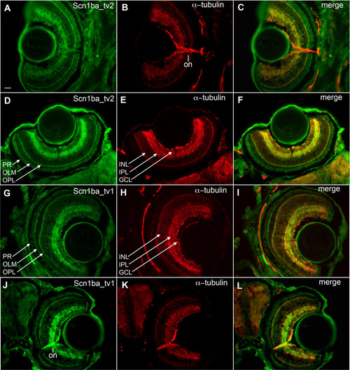
Retinal patterning of Scn1ba_tv1 and Scn1ba_tv2. A – F: anti-Scn1ba_tv2 (green), anti-acetylated α-tubulin (red). G – L: anti-Scn1ba_tv1 (green), anti-acetylated α-tubulin (red). Anti-Scn1ba_tv2 stains the layers of the retina, including the ganglion cell layer (GCL), inner plexiform layer (IPL), outer plexiform layer (OPL), outer limiting membrane (OLM), and photoreceptor cell layer (PR). Staining appears to be absent in the inner nuclear layer (INL) and in the optic nerve (on). Anti-Scn1ba_tv1 stains all the layers of the retina including the inner nuclear layer, where it shows robust staining. In contrast to anti-Scn1ba_tv2, anti-Scn1ba_tv1 labels optic nerve. Scale bar: 50 μm. EXPRESSION / LABELING:
|
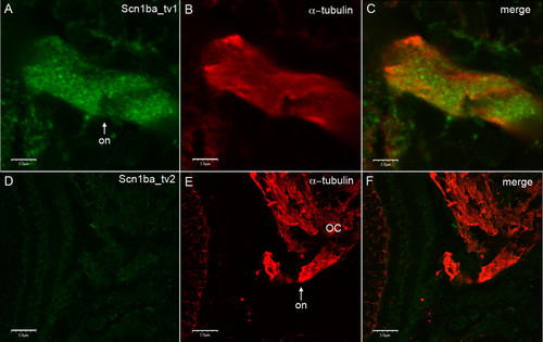
Zebrafish Scn1ba_tv1 but not Scn1ba_tv2 is expressed in optic nerve. Sections generated from 13 dpf zebrafish were stained with anti-Scn1ba_tv1 or anti-Scn1ba_tv2 and anti-acetylated α-tubulin. A – C: Anti Scn1ba_tv1 (green), anti-acetylated α-tubulin (red). D – F: Anti-Scn1ba_tv2 (green), anti-acetylated α-tubulin (red). Images were viewed with an Olympus FluoView 500 confocal microscope at 100× magnification with 5× additional zoom. Scale bar: 50 μm. EXPRESSION / LABELING:
|

Zebrafish Scn1ba_tv1 and Scn1ba_tv2 are differentially expressed in the spinal cord. A – C: Anti-Scn1ba_tv1 (green), anti-acetylated α-tubulin (red). D – F: Anti-Scn1ba_tv2 (green), anti-acetylated α-tubulin (red). SC: spinal cord. Scale bar: 50 μm. EXPRESSION / LABELING:
|
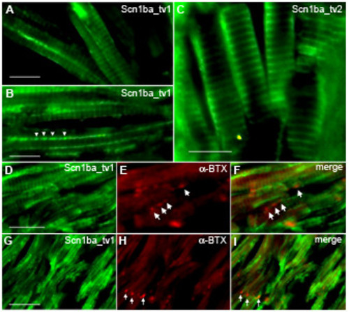
Zebrafish Scn1ba_tv1 and Scn1ba_tv2 are expressed in skeletal muscle. A, B, D, E, F: Anti-Scn1ba_tv1 β1 (green), α-bungarotoxin (BTX) (red). C. Anti-Scn1ba_tv2 (green). Labeling with anti-Scn1ba_tv1 produced two different staining patterns; staining at the t-tubules of striated muscle (A, D, and G), and punctate staining along the longitudinal edge of the muscle cells (arrowheads in B). Staining with anti-Scn1ba_tv2 labeled the t-tubule system and did not appear to label to muscle surface. Anti-Scn1ba_tv1 staining did not colocalize with α-BTX (D – F and H – I), suggesting that Scn1ba_tv1 is not expressed at neuromuscular junctions (arrows). Scale bar: 10 μm. EXPRESSION / LABELING:
|
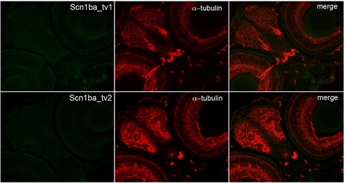
Antibody characterization. Immunohistochemical analysis of anti-Scn1ba_tv1 (top panel) or anti-Scn1ba_tv2 (lower panel) antibody (green) staining following pre-adsorption to its corresponding antigenic peptide. Sections were co-stained with anti-acetylated α-tubulin (red). Merged panels on the right. |