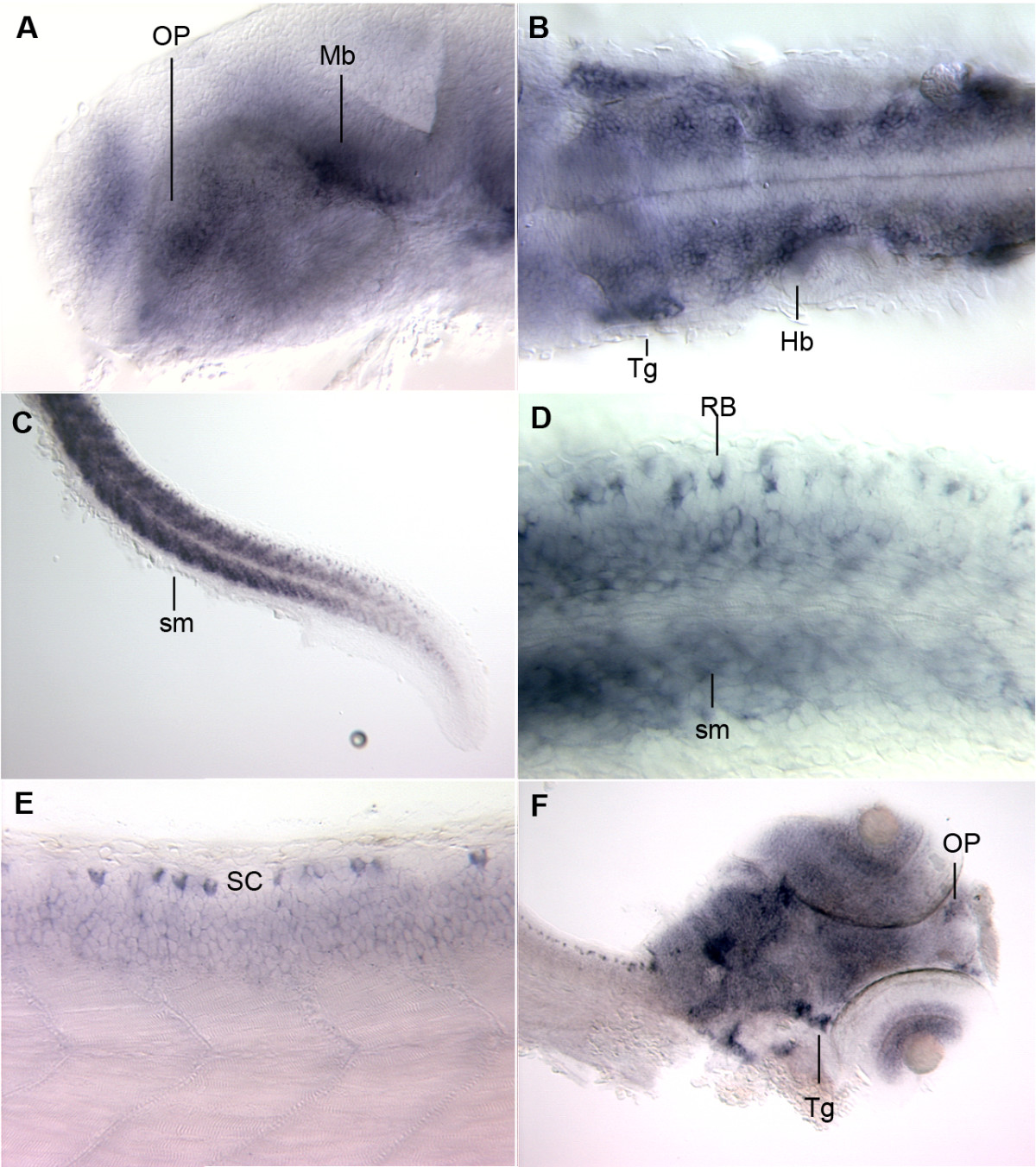
Fig. 2 In situ hybridization. A – D: Fish stained at 24 hpf. A. Staining is apparent in the olfactory placode (OP) and the midbrain (Mb). B. Dorsal mount showing staining in the trigeminal neuron (Tg) and in the rhombomeres of the hindbrain (Hb). C. Staining in spinal cord and skeletal muscle (sm). D. Higher magnification of Rohon Beard cells (RB) flanking skeletal muscle (sm). E. Fish at 48 hpf with staining in the Rohan Beard cells of the spinal cord (SC) and in the skeletal muscle. F. Staining throughout the brain, at the olfactory pits (OP), in the layers of the retina, and in the trigeminal ganglion (Tg) of fish at 72 hpf.
Image
Figure Caption
Figure Data
Acknowledgments
This image is the copyrighted work of the attributed author or publisher, and
ZFIN has permission only to display this image to its users.
Additional permissions should be obtained from the applicable author or publisher of the image.
Full text @ BMC Genomics

