
|
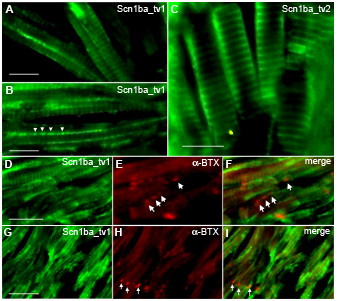
Fig. 10 Zebrafish Scn1ba_tv1 and Scn1ba_tv2 are expressed in skeletal muscle. A, B, D, E, F: Anti-Scn1ba_tv1 β1 (green), α-bungarotoxin (BTX) (red). C. Anti-Scn1ba_tv2 (green). Labeling with anti-Scn1ba_tv1 produced two different staining patterns; staining at the t-tubules of striated muscle (A, D, and G), and punctate staining along the longitudinal edge of the muscle cells (arrowheads in B). Staining with anti-Scn1ba_tv2 labeled the t-tubule system and did not appear to label to muscle surface. Anti-Scn1ba_tv1 staining did not colocalize with α-BTX (D – F and H – I), suggesting that Scn1ba_tv1 is not expressed at neuromuscular junctions (arrows). Scale bar: 10 μm.