
|
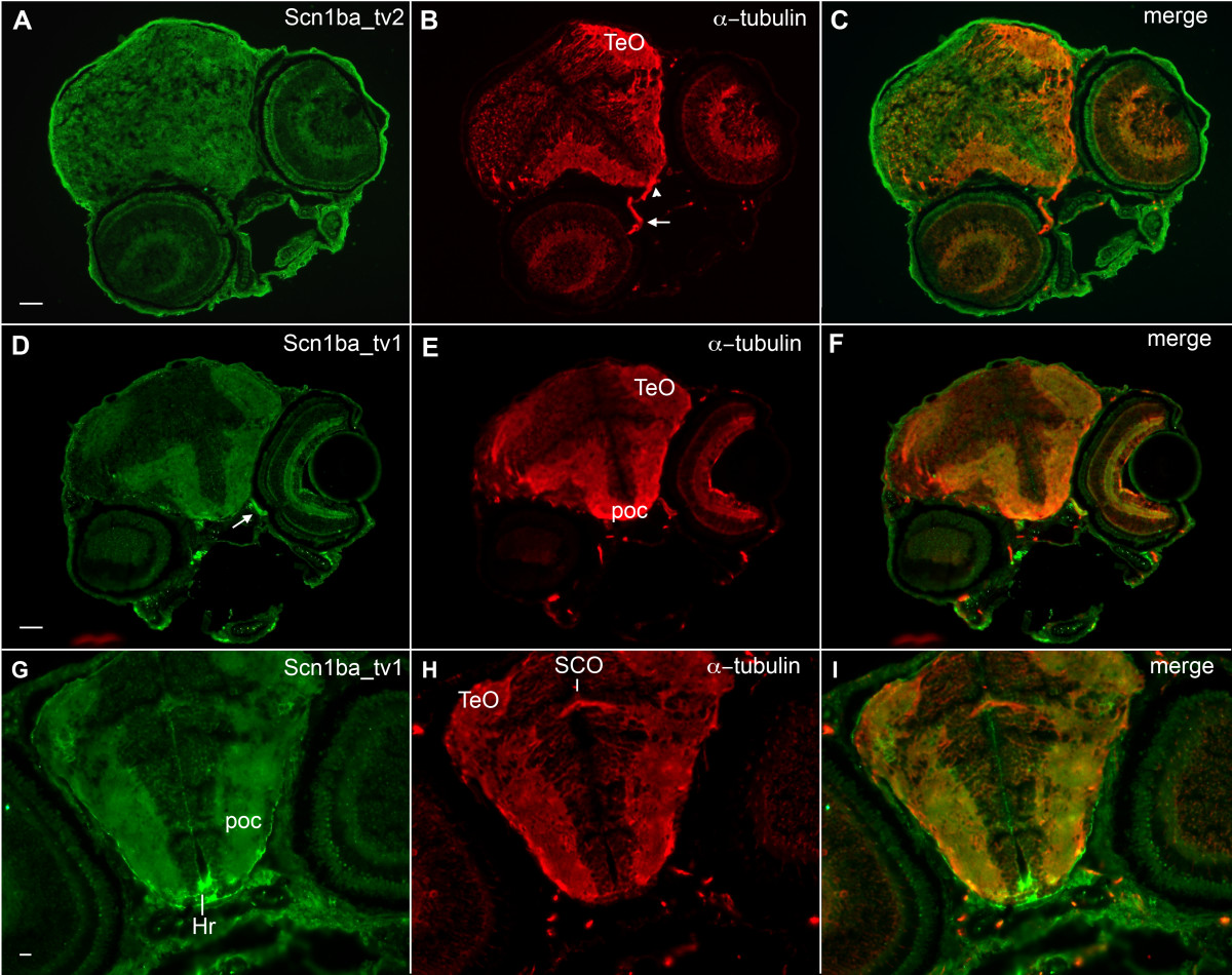
Fig. 6 Zebrafish Scn1ba_tv1 and Scn1ba_tv2 are expressed in brain. A – C. Anti-Scn1ba_tv2 (green), anti-acetylated α-tubulin (red). Arrow: optic nerve. Arrowhead: optic chiasm. Anti-Scn1ba_tv2 does not stain the optic nerve or optic chiasm. Scale bar: 20 μm. D – F. Anti-Scn1ba_tv1 (green), anti-acetylated α-tubulin (red). Scn1ba_tv1 staining appears in the optic tectum (TeO), post optic commissure (poc), and optic nerve (arrow). G – I. Anti-Scn1ba_tv1 (green), anti-acetylated α-tubulin (red). Scn1ba_tv1 staining appears in the poc and TeO as well as in the rostral hypothalamus (Hr), but is absent in the subcommisural organ (SCO).