- Title
-
Origin of the zebrafish endocrine and exocrine pancreas
- Authors
- Ward, A.B., Warga, R.M., and Prince, V.E.
- Source
- Full text @ Dev. Dyn.
|
Development of the pancreas in zebrafish and general fate map methodology. A: pdx1 is expressed bilaterally adjacent to the notochord of the anterior trunk at 14 hours postfertilization (hpf; based on Biemar et al.,[2001]). B-D: Morphogenesis of the pancreatic buds in zebrafish (based on Field et al.,[2003]). B: 24 hpf. The posterodorsal bud is visible. C: At 40 hpf. Both posterodorsal and anteroventral buds are visible, but have not fused. D: At 52 hpf. The buds have fused. E: One marginal blastomere was injected at the 512- to 2000-cell stage. F: Location of injected cells that gave rise to endoderm. Red circles were locations for the 40 hpf analysis, and blue squares were for the 72 hpf analysis. G-J: Embryos were imaged at various time points using brightfield and fluorescence, and photos were merged. G: At 6 hpf. H: At 10 hpf. I: At 24 hpf. J: Dorsal view of 72 hpf embryo showing rhodamine dextran-labeled cells in the exocrine and endocrine pancreas. Anti-Islet1 (blue) labels the endocrine pancreas and anti-green fluorescent protein (GFP; green) labels the postpharyngeal endoderm in gut-GFP embryos. DB, posterodorsal pancreatic bud; EP, exocrine pancreas; I, pancreatic islet; IB, intestinal bulb; L, liver; N, notochord; P, pancreas; S, embryonic shield; VB, anteroventral pancreatic bud. EXPRESSION / LABELING:
|
|
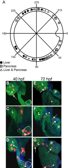
Fate map of liver and pancreatic progenitors at 6 hours postfertilization (hpf). A: Polar plot showing location at 6 hpf of progenitors of the liver and pancreas. The embryos from the 40 hpf and 72 hpf analyses were combined. Gray squares are pancreatic progenitors, black circles are liver progenitors, and white triangles indicate a mixed liver and pancreas population. B-G: All confocal images are individual slices from a Z-stack. B: Embryo with labeled cells in the liver bud at 40 hpf. C: Embryo with labeled cells in the pancreatic bud. D: Embryo with labeled cells in the liver and pancreatic buds at 40 hpf. E: Example of embryo with labeled cells only in the liver at 72 hpf. F: Embryo with labeled cells in the pancreas at 72 hpf. G: Embryo with rhodamine dextran-labeled cells in the liver and the pancreas at 72 hpf. Arrows point to labeled cells that colocalize with the structure of interest. Asterisks (*) point to rhodamine dextran-labeled cells that are not in the pancreas or liver. Dashed lines outline the anteroventral bud and solid lines outline the posterodorsal bud. L, liver; P, pancreas EXPRESSION / LABELING:
|
|
Fate map of the posterodorsal and anteroventral pancreatic buds. A: Polar plot of the locations of the posterodorsal and anteroventral pancreatic bud progenitors at 6 hours postfertilization (hpf). These points are the pancreas positive locations seen in Figure 2A. B-D: Confocal slices of representative 40 hpf embryos. B: Example of embryo with labeled cells in the anteroventral bud (purple circles in A). C: Embryo with rhodamine dextran-labeled cells in the posterodorsal bud (yellow squares in A). D: Embryo with labeled cells in both buds (blue triangles in A). Arrows point to labeled cells that colocalize with the structure of interest. Asterisks (*) point to rhodamine dextran-labeled cells that are not in the pancreas or liver. The dashed line encircles the location of the ventral bud, and the solid line encircles the location of the dorsal bud. EXPRESSION / LABELING:
|
|
Fate map of the endocrine and exocrine pancreas in zebrafish at 6 hours postfertilization (hpf). A: Polar plot of the locations of endocrine and exocrine pancreatic progenitors at mid-gastrulation. B: Graph of percentage exocrine cells at 72 hpf vs. dorsoventral location of the clone at 6 hpf. The numbers of exocrine and endocrine cells were counted, and the percentage exocrine is the number of exocrine cells divided by the total number of cells. C-E: Confocal slices of representative 72 hpf embryos. C: Embryo with rhodamine dextran-labeled cells in the exocrine pancreas. D: Embryo with labeled cells in the endocrine pancreas. E: Embryo with labeled cells in both the endocrine and exocrine pancreas. Blue cells are islet1 positive. Arrows point to labeled cells that colocalize with the structure of interest. Asterisks (*) point to rhodamine dextran-labeled cells that are not in the pancreas or liver. EP, exocrine pancreas; I, pancreatic islet; IB, intestinal bulb; L, liver. EXPRESSION / LABELING:
|
|
Location of cells at tail bud that potentially give rise to the pancreas. A: Lateral view of sox17 expression at tail bud. B: Dorsal view of sox17 expression at tail bud. n: notochord. C: Locations of all cells (excluding cells in the extreme anterior and posterior) from 20 different embryos that had one or more labeled cells in one or both of the pancreatic buds. Each color represents a different embryo. Embryos were mounted in dorsal view with the equator of the embryo (approximately the level of the first somite) closest to the viewer. The gray square marks an area that includes labeled cells from all embryos and marks the likely location of the pancreatic progenitors at the end of gastrulation (tail bud stage, 10 hours postfertilization [hpf]). The outline of the notochord is indicated by dashed lines. D-J: Representative confocal projections from time-lapse experiment between 95% epiboly and 14 somites (approximately 9.5 hpf-16 hpf). Images are approximately equally spaced in time. Red circle contains cells that later colocalize with pdx1-green fluorescent protein (GFP) positive cells. K: Location of rhodamine dextran-labeled cells (red) at 16 hpf with cells that are positive for GFP (green). The dashed yellow line is the future division between somites 1 and 2. The solid yellow line marks the boundary between somites 1 and 2. The solid white line marks the notochord. EXPRESSION / LABELING:
|


