- Title
-
Pancreas development in zebrafish: early dispersed appearance of endocrine hormone expressing cells and their convergence to form the definitive islet
- Authors
- Biemar, F., Argenton, F., Schmidtke, R., Epperlein, S., Peers, B., and Driever, W.
- Source
- Full text @ Dev. Biol.
|
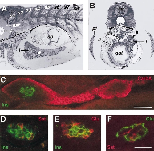
Morphology of the exocrine and endocrine pancreas in the zebrafish larva. (A) Sagittal and (B) cross sections of 6-day-old larvae showing the overall organization of the endocrine (i, arrow) and exocrine pancreatic tissues (e in A, and e, arrowhead in B). (C–F) Confocal images of pancreatic islet tissue showing whole-mount immunofluorescence staining performed on (C) 5.5-day-old, (D) 4-days, and (E, F) 3-day-old embryos with a combination of (C) anti-carboxypeptidase A and anti-insulin; (D) anti-insulin and anti-somatostatin; (E) anti-insulin and anti-glucagon; (F) anti-somatostatin and anti-glucagon antisera. In A and C, anterior is oriented to the left. da, dorsal aorta; e; exocrine tissue; i, islet; li, liver; o, otolith; pf, pectoral fin; sb, swim bladder; sc, spinal cord; s1–8, somite 1 to 8. Scale bar, 50 μm in C and 25 μm in F (same magnification as D and E). EXPRESSION / LABELING:
|
|
Sequence and expression of the zebrafish trypsin homolog. (A) Nucleotide and deduced amino acid sequences of the partial cDNA fragment. PCR primers used are underlined. (B, C) Dorsal view of 3- and 4-day-old embryos after in situ hybridization for trypsin. Anterior is to the left. EXPRESSION / LABELING:
|
|
Initiation of pdx-1 and insulin expression in the pancreatic primordium. (A–H) Dorsal and (I–N) lateral views of wild-type zebrafish embryos after in situ hybridization with pdx-1 (left panels) or insulin (right panels) antisense riboprobe at the (A, B) 10-somite stage; (C, D) 12-somite stage; (E, F) 14-somite stage; (G, H) 18-somite stage; (I, J) 20-somite stage; (K, L) 24-somite stage; and (M, N) 24 hpf. (O) 24 hpf embryo after in situ hybridization with both pdx-1 and insulin; n, notochord. The yolk was manually removed; anterior is to the left. Scale bar, 100 μm (N). EXPRESSION / LABELING:
|
|
Expression of somatostatin and glucagon in the developing zebrafish pancreas. Dorsal views of wild-type zebrafish embryos after in situ hybridization with somatostatin (left panels) or glucagon (right panels) antisense riboprobes at the (A, B) 16-somite stage; (C, D) 24-somite stage; and (E, F) 24 hpf. The yolk was manually removed; anterior is to the left. Scale bar, 100 μm. EXPRESSION / LABELING:
|
|
Pancreatic specific expression of the zebrafish nkx2.2, islet-1, and pax6.2 mRNAs. Whole-mount in situ hybridization of wild-type zebrafish embryos at the (A, A′) 10-somite stage; (C, C′, E, E′) 12-somite stage, and (B, D, F) 24 hpf performed with (A, A′, B) nkx2.2, (C, C′, D) islet-1, and (E, E′, F) pax6.2 antisense mRNA probes. Lateral views, dorsal to the right (A, A′, C, C′, E, E′) or to the top (B, D, F). Pancreas-specific expression domain are indicated (arrowheads). EXPRESSION / LABELING:
|
|
Development of the endocrine pancreas in zebrafish mutations affecting midline development and convergence movements. Expression of pdx-1 (A, C, E, G, I, K, M, O) and insulin (B, D, F, H, J, L, N, P) in wild-type and mutant embryos at 24 hpf. The following mutations are analyzed: (E, F) floating head; (G, H) one-eyed pinhead (oep); (M, N) spade tail (spt); and (O, P) knypek (kny). Lateral views in A, E, C, G, and dorsal views in all the other panels; anterior is to the left in all panels. All panels show the trunk of the embryo, between the posterior end of the hindbrain and about segment 10. The yolk has been removed from the embryos. |
Reprinted from Developmental Biology, 230(2), Biemar, F., Argenton, F., Schmidtke, R., Epperlein, S., Peers, B., and Driever, W., Pancreas development in zebrafish: early dispersed appearance of endocrine hormone expressing cells and their convergence to form the definitive islet, 189-203, Copyright (2001) with permission from Elsevier. Full text @ Dev. Biol.


