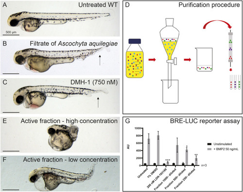
Identification of a fungal filtrate with activity consistentwith inhibition of BMP signaling. (A) Untreated control, not treated with fungal filtrate. WT, wild type. (B) An example of a phenotype induced by the filtrate of A. aquilegiae mixed in 1:1 ratio with E3 medium, with treatment at 6-48 hpf. (C) Phenotype induced by 750 nM DMH-1. The arrows in B and C indicate loss of the ventral fin. (D) Schematic overview of purification of active component. The filtrate is extracted with 3×1/3 volume ethyl acetate. The ethyl acetate fractions are than combined and dried. The residue is dissolved in DMSO and subsequently loaded onto a preparative HPLC column. Fractions are collected every 63 s. (E,F) Phenotypes induced by purified fraction, at high (E) and low (F) concentrations. In A-C,E,F, ten embryos were incubated per condition; representative pictures are shown. (G) Dose-dependent inhibitory effects of purified fraction of fungal filtrate on BMP2-induced Smad1/5-dependent BRE-luc transcriptional reporter activity. Lack of significant effect of vehicle control DMSO and potent antagonizing effect of BMPR type I kinase inhibitor LDN-193189 are shown. Results are expressed as mean±s.d., *P<0.05, **P<0.01, ***P<0.001 (Student’s t-test, two-tailed, unpaired).
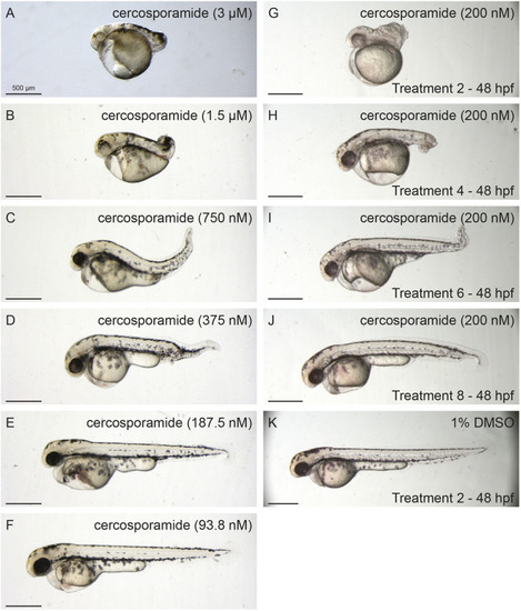
Dose- and time-dependent developmental defects of cercosporamide in zebrafish embryos. (A-F) Examples of phenotypes caused by a cercosporamide dilution range of 3 µM to 93 nM. (G-J) Examples of phenotypes caused by 200 nM cercosporamide with different treatment start times as indicated in hpf. (K) Control, 1% DMSO-treated embryo. Ten embryos were incubated per condition; representative pictures are shown.
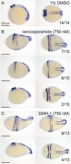
In situ hybridization using krox20/myod-specific probes confirms that cercosporamide and the well-known BMP inhibitor DMH-1 induced similar defects in zebrafish development. (A-C) Embryos were treated with 1% DMSO (control) (A), 750 nM cercosporamide (B) or 750 nM DMH-1 (C) from 6 hpf onwards and fixed at 12 hpf. In situ hybridization was performed using krox20/myod-specific probes. Representative examples of resulting embryos are shown with lateral view on the left and dorsal view on the right. In the bottom-right corner, the fraction of embryos showing the pattern is depicted.
Cercosporamide and known BMP inhibitors cooperate. Combination treatments of zebrafish embryos suggest that cercosporamide acts on the BMP signaling pathway. Embryos were treated with cercosporamide (50 nM or 100 nM), LDN-193189 (5 µM or 10 µM) or combinations from 7 hpf onwards as indicated. Ten embryos were incubated per condition; representative pictures of treated embryos are shown.
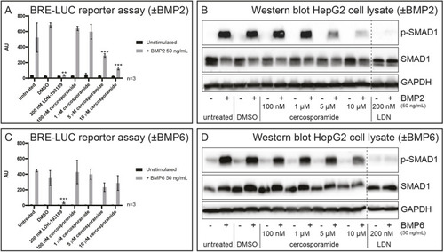
Cercosporamide inhibits BMP/SMAD signaling in mammalian cells. (A) HepG2 cells with BRE-luc reporter were treated with BMP2 (50 ng/ml) or not treated (−). Control (1% DMSO), LDN-193189 (200 nM) or a range of concentrations of cercosporamide (100 nM to 10 µM) were added and luciferase activity was determined. Averages of triplicate measurements are depicted as arbitrary units. (B) HepG2 cells were treated with BMP2 (50 ng/ml) or not (−), and with 1% DMSO (control), a range of concentrations of cercosporamide (100 nM to 10 µM) or LDN-193189 (200 nM). Cells were lysed and the lysates run on SDS-PAGE gels. The material on the gel was transferred to blots and parallel blots were probed using antibodies, specific for phosphoSMAD1/5/8 (p-SMAD1) and SMAD1 or GAPDH (loading control). Detection was performed by enhanced chemiluminescence (ECL). Representative blots are shown. (C) As in A, except BMP6 (50 ng/ml) was used instead of BMP2. (D) As in B, except BMP6 (50 ng/ml) was used instead of BMP2. **P<0.01 and ***P<0.001 (Student’s t-test, two-tailed, unpaired). All samples in B and D were run on the same gel; the dashed lines indicate where the blots were cut.
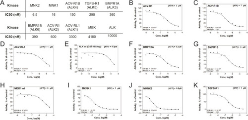
Cercosporamide inhibits kinase activity of purified ALK receptors in vitro. (A) IC50 values of cercosporamide-mediated inhibition of a panel of ten kinases. (B-K) Activity graphs from which the IC50 values in A were derived. The kinases include six type I ALK BMPRs, mitogen-activated protein kinase (MAPK)-interacting protein kinases 1 and 2 (MNK1, MNK2), mitogen-activated protein kinase kinase (MEK) and the tyrosine kinase, anaplastic lymphoma kinase (ALK). wt, wild type.
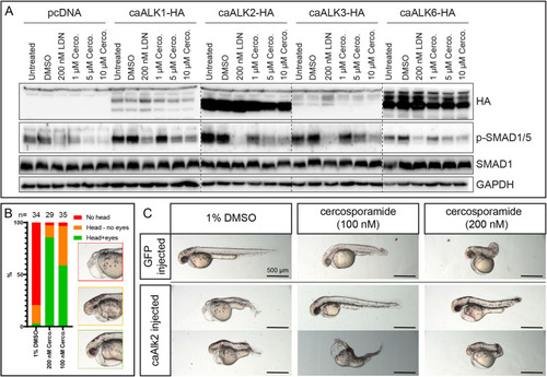
Cercosporamide selectively inhibits caBMPR type I in cultured mammalian cells and zebrafish embryos. (A) HEK 293T cells were transfected with empty vector (pcDNA) or expression vectors for caALK1, caALK2, caALK3 or caALK6 each with a haemagglutinin (HA) epitope tag at the carboxy terminus. The cells were treated with vehicle (1% DMSO), LDN193189 (200 nM) or a range of cercosporamide concentrations (1-10 µM as indicated). Subsequently, the cells were lysed and the lysates run on SDS-PAGE gels. The material on the gel was transferred to blots and parallel blots were probed with antibodies specific for HA (epitope tag on receptors), phosphoSMAD1/5/8 (p-SMAD1), SMAD1 or GAPDH (loading control). Dashed lines indicate the borders of different gels. (B) Cercosporamide partially rescued caAlk2-induced developmental defects in zebrafish embryos in vivo. Bar chart shows the phenotype distribution of embryos injected with caAlk2 mRNA and subsequently treated with 1% DMSO, 100 nM or 200 nM cercosporamide from 2 hpf onwards. The severity of the phenotype (examples depicted in the insets) is plotted as red (severe; no head), orange (intermediate; head structures present, no eyes) and green (mild; head structure with eyes detectable). The total number of embryos (n) is indicated. (C) The phenotypes of the rescued embryos are highly variable; therefore, we depicted two representative individuals of ten embryos that were treated for each condition.
Acknowledgments
This image is the copyrighted work of the attributed author or publisher, and
ZFIN has permission only to display this image to its users.
Additional permissions should be obtained from the applicable author or publisher of the image.
Full text @ Dis. Model. Mech.
Your Input Welcome
Thank you for submitting comments. Your input has been emailed to ZFIN curators who may contact you if
additional information is required.
Oops. Something went wrong. Please try again later.