IMAGE

Fig. 4.

ID
ZDB-IMAGE-201003-120
Source
Figures for Hoeksma et al., 2020
Image
Figure Caption

Fig. 4.

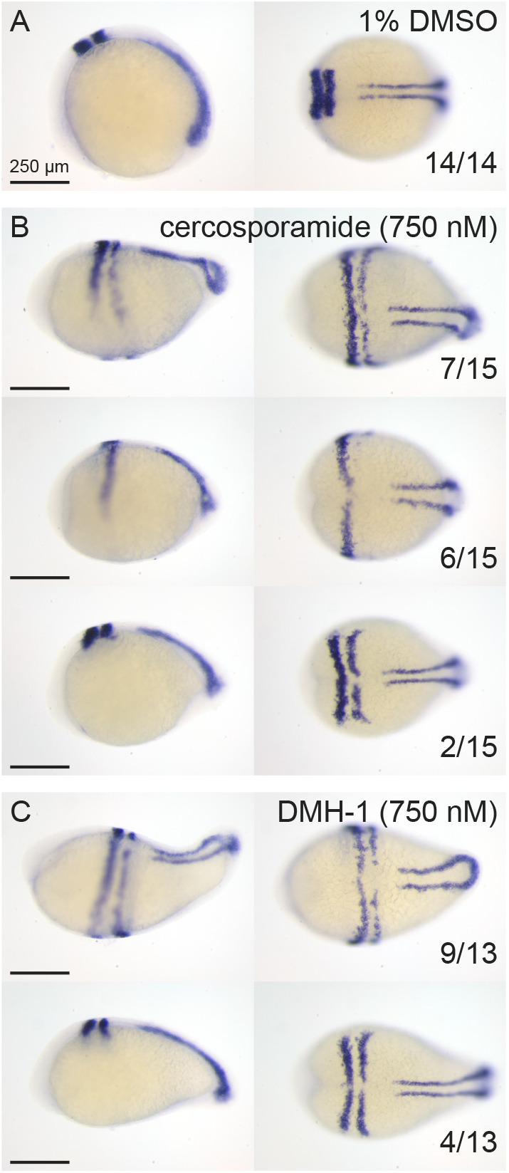
In situ hybridization using krox20/myod-specific probes confirms that cercosporamide and the well-known BMP inhibitor DMH-1 induced similar defects in zebrafish development. (A-C) Embryos were treated with 1% DMSO (control) (A), 750 nM cercosporamide (B) or 750 nM DMH-1 (C) from 6 hpf onwards and fixed at 12 hpf. In situ hybridization was performed using krox20/myod-specific probes. Representative examples of resulting embryos are shown with lateral view on the left and dorsal view on the right. In the bottom-right corner, the fraction of embryos showing the pattern is depicted.

Acknowledgments
This image is the copyrighted work of the attributed author or publisher, and ZFIN has permission only to display this image to its users. Additional permissions should be obtained from the applicable author or publisher of the image. Full text @ Dis. Model. Mech.