IMAGE

Fig. 6.

ID
ZDB-IMAGE-201003-121
Source
Figures for Hoeksma et al., 2020
Image
Figure Caption

Fig. 6.

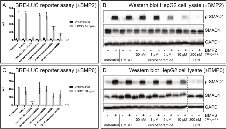
Cercosporamide inhibits BMP/SMAD signaling in mammalian cells. (A) HepG2 cells with BRE-luc reporter were treated with BMP2 (50 ng/ml) or not treated (−). Control (1% DMSO), LDN-193189 (200 nM) or a range of concentrations of cercosporamide (100 nM to 10 µM) were added and luciferase activity was determined. Averages of triplicate measurements are depicted as arbitrary units. (B) HepG2 cells were treated with BMP2 (50 ng/ml) or not (−), and with 1% DMSO (control), a range of concentrations of cercosporamide (100 nM to 10 µM) or LDN-193189 (200 nM). Cells were lysed and the lysates run on SDS-PAGE gels. The material on the gel was transferred to blots and parallel blots were probed using antibodies, specific for phosphoSMAD1/5/8 (p-SMAD1) and SMAD1 or GAPDH (loading control). Detection was performed by enhanced chemiluminescence (ECL). Representative blots are shown. (C) As in A, except BMP6 (50 ng/ml) was used instead of BMP2. (D) As in B, except BMP6 (50 ng/ml) was used instead of BMP2. **P<0.01 and ***P<0.001 (Student’s t-test, two-tailed, unpaired). All samples in B and D were run on the same gel; the dashed lines indicate where the blots were cut.

Acknowledgments
This image is the copyrighted work of the attributed author or publisher, and ZFIN has permission only to display this image to its users. Additional permissions should be obtained from the applicable author or publisher of the image. Full text @ Dis. Model. Mech.