IMAGE

Fig. 8.

ID
ZDB-IMAGE-201003-124
Source
Figures for Hoeksma et al., 2020
Image
Figure Caption

Fig. 8.

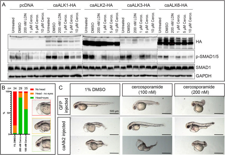
Cercosporamide selectively inhibits caBMPR type I in cultured mammalian cells and zebrafish embryos. (A) HEK 293T cells were transfected with empty vector (pcDNA) or expression vectors for caALK1, caALK2, caALK3 or caALK6 each with a haemagglutinin (HA) epitope tag at the carboxy terminus. The cells were treated with vehicle (1% DMSO), LDN193189 (200 nM) or a range of cercosporamide concentrations (1-10 µM as indicated). Subsequently, the cells were lysed and the lysates run on SDS-PAGE gels. The material on the gel was transferred to blots and parallel blots were probed with antibodies specific for HA (epitope tag on receptors), phosphoSMAD1/5/8 (p-SMAD1), SMAD1 or GAPDH (loading control). Dashed lines indicate the borders of different gels. (B) Cercosporamide partially rescued caAlk2-induced developmental defects in zebrafish embryos in vivo. Bar chart shows the phenotype distribution of embryos injected with caAlk2 mRNA and subsequently treated with 1% DMSO, 100 nM or 200 nM cercosporamide from 2 hpf onwards. The severity of the phenotype (examples depicted in the insets) is plotted as red (severe; no head), orange (intermediate; head structures present, no eyes) and green (mild; head structure with eyes detectable). The total number of embryos (n) is indicated. (C) The phenotypes of the rescued embryos are highly variable; therefore, we depicted two representative individuals of ten embryos that were treated for each condition.

Acknowledgments
This image is the copyrighted work of the attributed author or publisher, and ZFIN has permission only to display this image to its users. Additional permissions should be obtained from the applicable author or publisher of the image. Full text @ Dis. Model. Mech.