
- Title
-
A zebrafish sox9 gene required for cartilage morphogenesis
- Authors
- Yan, Y.-L., Miller, C.T., Nissen, R.M., Singer, A., Liu, D., Kirn, A., Draper, B., Willoughby, J., Morcos, P.A., Amsterdam, A., Chung, B.-C., Westerfield, M., Haffter, P., Hopkins, N., Kimmel, C., and Postlethwait, J.H.
- Source
- Full text @ Development
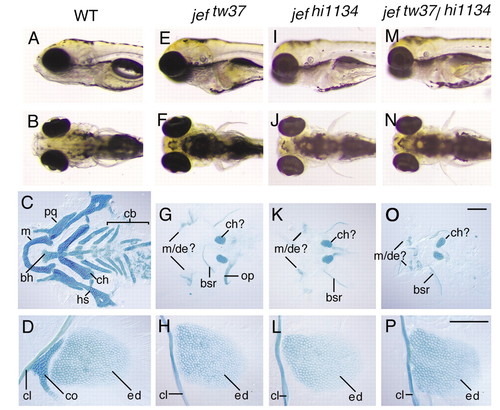
Activity of jef is essential for development of many cartilages. (A,E,I,M, lateral views, anterior towards the left; B,F,J,N, ventral views, anterior towards the left; C,G,K,O, dissected pharyngeal cartilages stained with Alcian, anterior towards the left; D,H,L,P, Alcian stained right pectoral and fin skeletons, anterior towards the top. (A-D) Wild type; (E-H) homozygous jeftw37; (I-L) homozygous jefhi1134; (M-P) heterozygous jeftw37/jefhi1134. All animals are 5 dpf. bh, basihyal; bsr, branchiostegal rays; cb, ceratobranchials; ch, ceratohyal; ch?, presumed ceratohyal; cl, cleithrum; ed, endoskeletal disk; co, scapulocoracoid; hs, hyosymplectic; m, Meckel?s cartilage; m/de? putative Meckel?s cartilage and dentary bone; op, opercule; pq, palatoquadrate. Scale bars: 100 μm in O for C,G,K,O; 100 μm in P for D,H,L,P. PHENOTYPE:
|
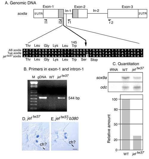
jeftw37 alters a conserved splice site sequence in sox9a and inhibits message splicing. (A) The sequence of sox9a in zebrafish wild-type strains AB and T�, splice junction (arrow). An in frame stop codon directly followed the G to T change in jeftw37. (B) The primer pair f1/r1 in exon 1 and intron 1 amplified a band of the expected size (544 bp) from wild-type (WT) genomic DNA and from jeftw37 cDNA, showing that intronic sequences of sox9a were present in mutant cDNA. With normal splicing in wild-type cDNA, there was no strong product of this size. (C) RNase protection assays (RPA, upper panel) on RNA extracted from wild-type and homozygous jeftw37 mutants 4 days old. tRNA served as a negative control. odc, the internal control. Probe for RPA is made from a 402 bp of sox9a cDNA amplified using primer pair f2/r2. Quantifying band intensity (lower panel) shows a drastic decrease of message in jeftw37 embryos. (D,E) The jeftw37 allele behaves as a null when heterozygous for the deletion allele b380. bh, basihyal; bsr, branchiostegal rays; cb, ceratobranchials; ch, ceratohyal; ch?, presumed ceratohyal; cl, cleithrum; ed, endoskeletal disk; co, scapulocoracoid; hs, hyosymplectic; m, Meckel?s cartilage; m/de? putative Meckel?s cartilage and dentary bone; op, opercule; pq, palatoquadrate. Scale bar: 100 μm. |
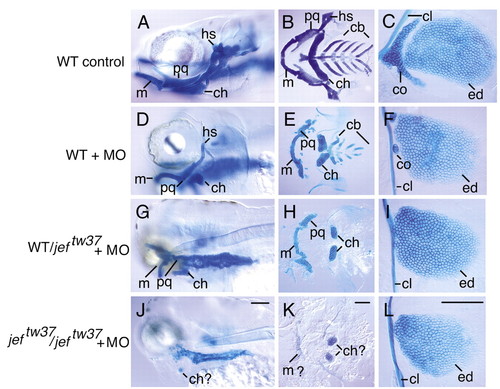
Splice-directed morpholinos to sox9a produce a phenotype similar to jef. (A-C) Uninjected wild-type controls. (D-F) Homozygous wild types injected with MO i1d plus i2d. (G-I) Heterozygotes for jeftw37 injected with MOs. (J-L) MO injected homozygous jeftw37 individuals. (A,D,G,J) Lateral view of 5 dpf animals, anterior towards the left. (B,E,H,K) Ventral view of dissected cartilages, anterior towards the left. (C,F,I,L) Right pectoral and fin skeleton, proximal towards the left. bh, basihyal; bsr, branchiostegal rays; cb, ceratobranchials; ch, ceratohyal; ch?, presumed ceratohyal; cl, cleithrum; ed, endoskeletal disk; co, scapulocoracoid; hs, hyosymplectic; m, Meckel?s cartilage; m/de? putative Meckel?s cartilage and dentary bone; op, opercule; pq, palatoquadrate. Scale bars: in J, 100 μm for A,D,G,J; in K, 100 μm for B,E,H,K; in L, 100 μm for C,F,I,L. PHENOTYPE:
|
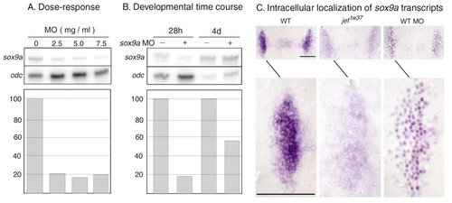
Morpholino antisense oligonucleotides directed against sox9a inhibit splicing and transport of sox9a transcript. (A) RNase protection assays of sox9a transcript in 28 hpf wild-type animals injected at the one-cell stage with 2 nl of different concentrations of MO, normalized against odc. (B) RNase protection assays of sox9a transcript in animals 28 hpf or 4 dpf either treated (+) or not treated (?) with 5.0 ng MO, normalized against odc. Graphs show the amount of RNA present as a percent of untreated controls. (C) Transcript for sox9a accumulates in the cytoplasm in wild type, but in the nucleus in splice-directed MO injected embryos at 28 hpf. In jeftw37 embryos, transcript is visible both in the cytoplasm and in the nucleus. Dorsal views, anterior is upwards. Equal amounts of MO directed against the donor sites for intron 1 and 2. Scale bars: 100 μm. EXPRESSION / LABELING:
|
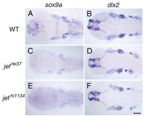
Activity of sox9a is not required for the specification or migration of cranial neural crest. (A,B) Wild type (WT) embryos. (C,D) Homozygous jeftw37 embryos. (E,F) Homozygous jefhi1134 embryos. (A,C,E) sox9a expression. (B,D,F) dlx2 expression. All animals were 24 hpf. Dorsal views, anterior towards the left. EXPRESSION / LABELING:
|
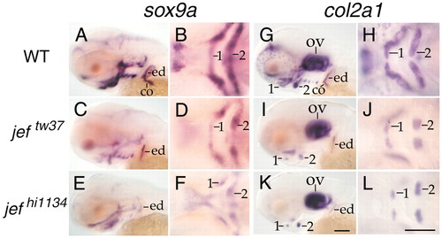
Activity of sox9a is necessary for the expression of col2a1 in most developing cartilage of the neurocranial, pharyngeal and pectoral skeleton. (A-F) Expression of sox9a. (G-L) Expression of col2a1. (A,B,G,H) Wildtype animals. (C,D,I,J) Homozygous mutant jeftw37 embryos. (E,F,K,L) Homozygous mutant jefhi1134 embryos. Photographs are a montage with focus on the pharyngeal cartilages, and the pectoral girdle. All animals are 68 hpf. 1 and 2, first and second pharyngeal arches; co, scapulocoracoid; ed, endoskeletal disc; ov, otic vesicle. (A,C,E,G,I,K) Lateral views, anterior towards the left. (B,D,F,H,J,L) Ventral views, anterior towards the left. Scale bars: in K, 100 mm for A,C,E,G,I,K; in L, 100 μm for B,D,F,H,J,L. EXPRESSION / LABELING:
|
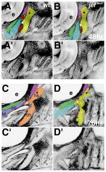
Prechondrogenic condensations form in jef (sox9a) mutants, but differentiation and morphogenesis fail to occur. Confocal micrographs (lateral views shown as negative images, anterior towards the left) of live wild-type (A,A′,C,C′) and jeftw37 mutants (B,B′,D,D′), stained with BODIPY-ceramide. Cells appear white and interstitial space appears black. (The pharyngeal cavity does not retain the dye, so it also appears white in C-D′). At 48 hours, in wild types (A,A′) and jef mutants for both alleles (B,B′, and data not shown), prechondrogenic condensations have formed. The original images (A′,B′) are pseudo-colored (A,B) to highlight the first (blue) and second (yellow) pharyngeal arch precartilage condensations. Red, first aortic arch; green, adductor mandibulae muscle; pink, constrictor dorsalis premyogenic condensation. At this stage, a contiguous condensation prefigures the dorsal and ventral pharyngeal cartilages, as seen best for the second arch in this focal plane (A-B′). In the second arch precartilage condensation, the symplectic rudiment (white asterisk) and hyomandibular foramen (black asterisk) are apparent in wild types and jef mutants (A-B′). By 76 hours, individuated and precisely shaped dorsal and ventral first and second pharyngeal arch cartilages with highly ordered stacks of chondrocytes have formed in wild types (C,C�). Original images (C′,D′) are pseudocolored (C,D as in A,B) with overtly differentiated cartilage in wild types colored purple in the first arch and orange in the second arch cartilages. Stacking, individuation, and proper shaping do not occur in jef mutants (D,D′). The precise boundary of the first and second arch is not visible in jef mutants and is approximated in the pseudocolored panel (D). For jeftw37, five mutants and 15 phenotypically wild-type siblings were examined; for jefhi1134, nine mutants and 21 phenotypically wild-type siblings were examined. ch, ceratohyal; e, eye; hs, hyosymplectic; M, Meckel?s; pq, palatoquadrate (see Fig. 1). |
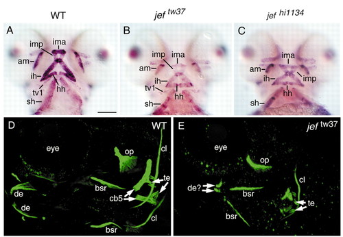
Muscle and bone in jef mutants. Expression of titin in wild-type (A) and homozygous jeftw37 (B) and jefhi1134 (C) animals at 3 dpf reveals normal patterning of cranial muscles. Compiled stacks of confocal micrographs of wildtype (D) and a jeftw37 mutant (E) stained with calcein. Most dermal bones (op, bsr, cl) are relatively unaffected in jef mutants, whereas the dermal dentary (de) and cartilage replacement fifth ceratobranchial bone (cb5) are severely reduced. Teeth (te) are present, as are tiny remnants of bone at the base of the teeth. Bone development occurs on schedule without enlarged ossification centers in jef mutants. am, adductor mandibulae; bsr, branchiostegal ray; cb5, fifth ceratobranchial; cl, cleithrum; de, dentary; hh, hyohyoideus; ih, interhyoideus; ima, intermandibularis anterior; imp, intermandibularis posterior; op, opercle; sh, sternohyoideus; te, teeth; tv1, transversus ventralis. Scale bar: in A, 100 μm for A-C. EXPRESSION / LABELING:
|