
IMAGE

Fig. 3.

ID
ZDB-IMAGE-260311-65
Genes
Publication
Tarbashevich et al., 2026 - Cell-autonomous control coupled with tissue context regulates the cessation of migration at the site of organ development
All Figures
Figures for Tarbashevich et al., 2026
Image
Figure Caption

Fig. 3.

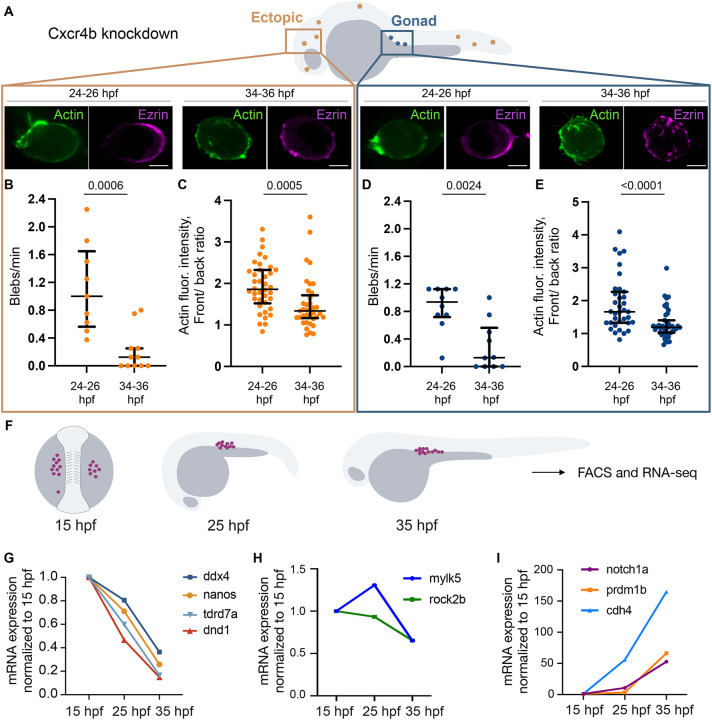
Cellular events during motility loss. (A) PGCs at gonad region and ectopic positions were examined for blebbing activity and polarity. Localization of Lifeact and Ezrin in ectopic and gonadal PGCs. Scale bars: 5 µm. (B,D) Blebbing frequency in ectopic PGCs (B) and in PGCs at the gonad (D). Ectopic PGCs: 9 (24-26 hpf), 11 (34-36 hpf); gonadal PGCs: 10 (24-26 hpf), 10 (34-36 hpf). (C,E) Quantification of the PGC polarity based on the cell front/back ratio for the Lifeact fluorescence. Ectopic PGCs: 38 (24-26 hpf), 37 (34-36 hpf); gonadal PGCs: 35 (24-26 hpf), 43 (34-36 hpf). For B-E, Mann–Whitney test. Data are median±error bars (IQR). (F) Embryos at 15-35 hpf were used for FACS of PGCs (magenta) and sequencing. (G-I) Normalized levels of mRNAs important for PGC development (G), contractility (H) and adhesion/differentiation (I).

Figure Data
Acknowledgments
This image is the copyrighted work of the attributed author or publisher, and ZFIN has permission only to display this image to its users. Additional permissions should be obtained from the applicable author or publisher of the image. Full text @ Development