
Fig. 2
- ID
- ZDB-IMAGE-201005-13
- Genes
- Publication
- Ogawa et al., 2019 - Six6 and Six7 coordinately regulate expression of middle-wavelength opsins in zebrafish
- All Figures
- Figures for Ogawa et al., 2019
|
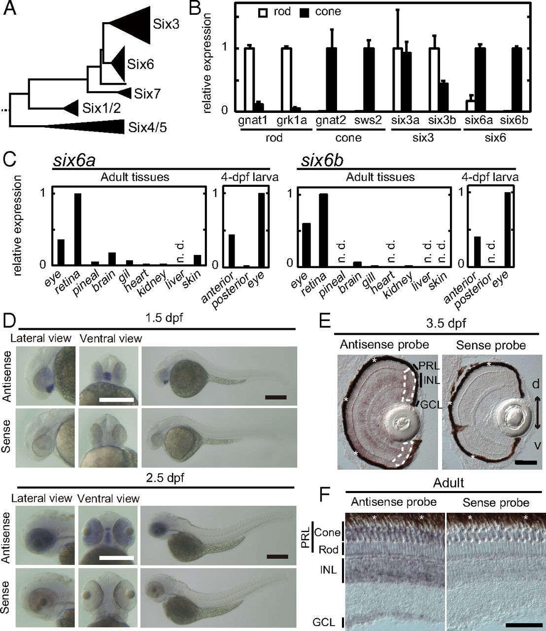
Fig. 2 Expression pattern of six6a and six6b. (A) Schematic representation of the phylogenetic tree of the Six family, modified from our previous study (17). Relative expression levels of six6a and six6b are shown in isolated rods and cones at the adult stage (mean ± SD, n = 2) (B) and in adult and 4-dpf larval tissues (C). n.d., not detected. Photoreceptor purity after isolation was further validated in SI Appendix, Fig. S1. Expression patterns of six6b examined by in situ hybridization in whole-mount embryos at 1.5 dpf and 2.5 dpf (D), in the larval retina at 3.5 dpf (E), and in the 6-mo postfertilization adult retina (F) are shown. In E, ciliary marginal zones are surrounded by white broken lines. d, dorsal side; GCL, ganglion cell layer; INL, inner nuclear layer; PRL, photoreceptor layer; v, ventral side. The retinal pigmented epithelium (indicated by asterisks) is adjacent to the photoreceptor layer. (Scale bars: D, 300 μm; E and F, 50 μm.)