
Fig. 2
|
Fig. 2
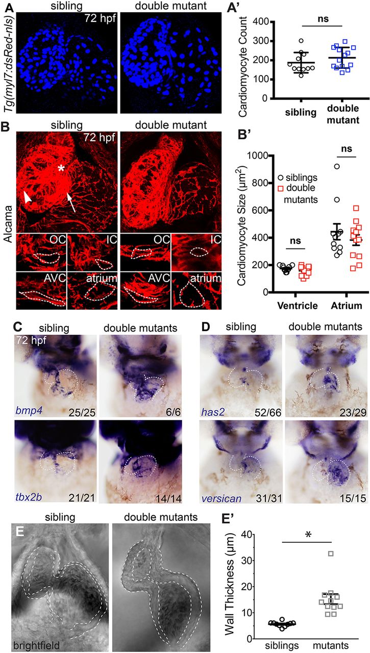
AVC markers are expanded in double mutant embryos and cardiac jelly is thicker. (A) Confocal z-stacks of Tg(myl7:dsRed-nls) embryos (labelling cardiomyocyte nuclei) show no difference in cell number between sibling and double mutant embryos. (A′) Graphical representation of cell number (n=11 siblings; n=14 mutants). (B) Confocal images of representative sibling and double mutant embryos immunostained for Alcama to visualise cell shape. IC, inner curvature (white asterisk); OC, outer curvature (white arrowhead); AVC, atrioventricular canal (white arrow). Individual cardiomyocytes are outlined with broken lines. (B′) Graph showing average cardiomyocyte size of ventricle and atrium in siblings and double mutants (n=11 siblings; n=12 mutants). (C) In situ hybridisation analysis of AVC markers bmp4, tbx2b, has2 and versican are expanded into the atrial chamber of double mutants compared with siblings. (E) Bright-field still images from high-speed movies of hearts at atrial diastole show thickened space (outlined) between the myocardial outer surface and the lumen surface of the heart, indicating thicker cardiac jelly in double mutants. (E′) Quantification of cardiac jelly thickness showing a significant increase in double mutant embryos compared with wild-type siblings when measuring the outer atrial myocardial wall and the inner luminal lining (n=10 siblings; n=11 mutants). ns, not significant; *P<0.05, as determined by t-test. Data are mean±s.d.