Image
Figure Caption

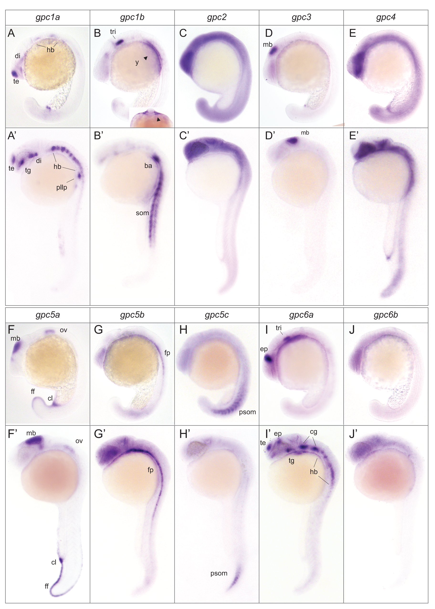
Fig. 6 Spatial expression pattern of zebrafish glypicans during segmentation and pharyngula stages.

In situ hybridization was carried out to detect glypican mRNA at 20 somite stage: 19 hpf and pharyngula stages: 24-26 hpf. Lateral view of whole mount embryos, anterior to the left. Expression pattern of gpc1a (A, A′), gpc1b (B, B′); subfigure in B is an optical section indicating expression in yolk cells (arrowheads) and branchial arch primordium, gpc2 (C, C′), gpc3 (D, D′), gpc4 (E, E′), gpc5a (F, F′), gpc5b (G, G′), gpc5c (H, H′), gpc6a (I, I′), gpc6b (J, J′). di, diencephalon; te, telencephalon; hb, hindbrain; tg, tegmentum; nc, neural crest; pm, paraxial mesoderm; som, somites; ba, branchial arch; y, yolk; mb, midbrain; ff, fin fold; ov, otic vesicle; fp, floor plate; psom, posterior somites; ep, epiphysis; tri, trigeminal placode; cl, cloaca; pllp, posterior lateral line primordium; cg, cranial ganglion.

Figure Data
Acknowledgments
This image is the copyrighted work of the attributed author or publisher, and ZFIN has permission only to display this image to its users. Additional permissions should be obtained from the applicable author or publisher of the image. Full text @ PLoS One