
Fig. 1
- ID
- ZDB-IMAGE-140319-43
- Genes
- Publication
- Nguyen et al., 2000 - Dorsal and intermediate neuronal cell types of the spinal cord are established by a BMP signaling pathway
- All Figures
- Figures for Nguyen et al., 2000
|
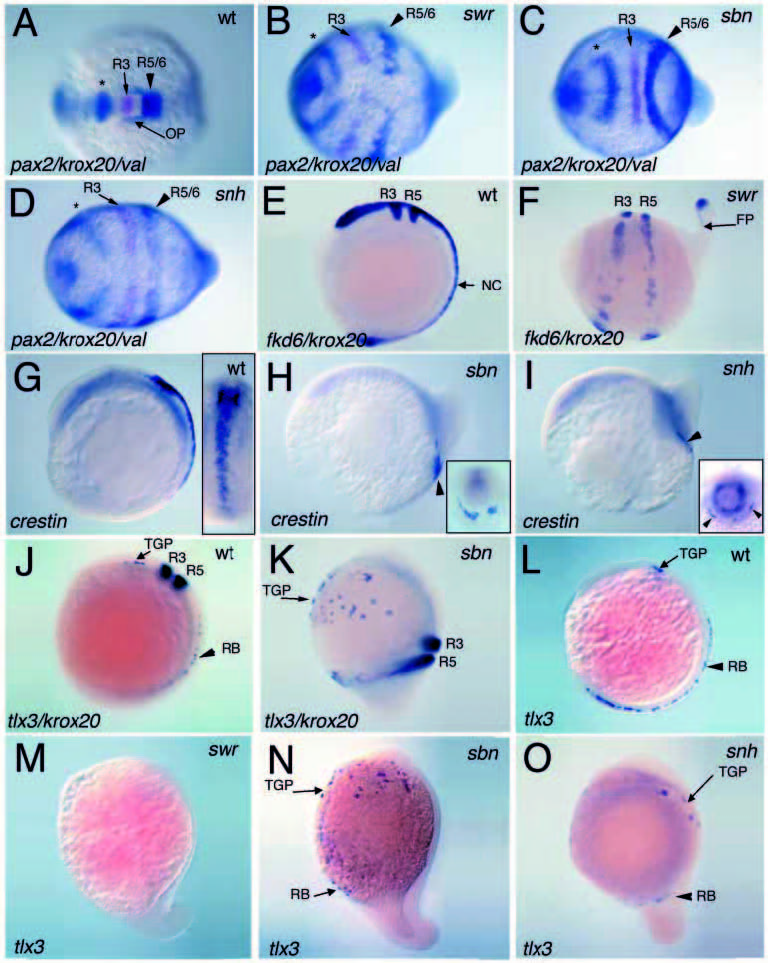
Fig. 1 Conserved anteroposterior neural patterning (A-D) and loss of trunk neural crest (E-I) and dorsal RB neurons (J-O) in dorsalized mutant embryos. (A-D) are two-color triple in situ hybridizations in 6- somite-stage wild-type (A), swr/bmp2b (B), sbn/smad5 (C) and snh/bmp7 (D) embryos (dorsal views with anterior to the left). Expression of pax2.1 in the midbrain-hindbrain boundary (marked with an *) and val in rhombomeres 5 and 6 are in blue, while krox20 expression in rhombomere 3 is in magenta. Similar results were found in 1- to 2-somitestage embryos. Expression of fkd6 and krox20 in wild-type (E) and a swr/bmp2b mutant (F) at the 6- somite stage. crestin expression posterior to rhombomere 5 and within the rostral trunk of a 9-somitestage wild-type embryo (G), and in a greatly reduced domain in sbn/smad5 (H) and snh/bmp7 (I) mutant embryos. Insets in G-I are dorsal or posterior views. Expression of tlx3 and krox20 in 7-somite-stage wildtype (J) and sbn/smad5 (K) embryos. tlx3 expression in 7-somite wild-type (L), swr/bmp2b (M), sbn/smad5 (N) and snh/bmp7 (O) mutant embryos. In 0/8 swr/bmp2b, 8/20 sbn/smad5 and 5/8 snh/bmp7 mutant embryos, some RB-tlx3 expressing cells were detected, but in severely reduced numbers. Lateral views (E-O) with anterior to the top, dorsal to the right in E,G,J-O, and anterior to the left, dorsal to the top in F,H,I. R3, rhombomere 3; R5, rhombomere 5; R5/6, rhombomeres 5 and 6; OP, otic placode; NC, neural crest; FP, floor plate; TGP, trigeminal placode precursors.