
Fig. 10
- ID
- ZDB-IMAGE-130530-3
- Genes
- Publication
- Fernandez et al., 2013 - Fixation/permeabilization: New alternative procedure for immunofluorescence and mRNA in situ hybridization of vertebrate and invertebrate embryos
- All Figures
- Figures for Fernandez et al., 2013
|
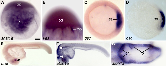
Fig. 10 mRNA in situ hybridization of whole-mounted zebrafish zygotes (A,B), gastrulae (C,D), and 1-day-old (E–G) larvae [Form-Acet]. A: Distribution of snai1a mRNA in the blastodisc (bd) and endoplasmic lacunae (el). B: Distribution of vas mRNA in meridional streamers (ms) and base of the blastodisc (bd). C: Expression of gsc mRNA in cells of the embryonic shield (es). D: Control showing the expression of gsc mRNA in cells of the embryonic shield (es) using the Thisse and Thisse (2008) protocol. Differences between the quality of the in situ hybridizations shown in C and D are confirmed when we compare our results with those obtained by others with a similar method in the same embryonic material (see Shao et al., 2012). E: Expression of brul mRNA in the lens (arrowhead). F: Expression of atoh1a mRNA in the hindbrain (arrowheads). G: Dorsal view of the hindbrain showing expression of atoh1a in the developing cerebellum (cr) and rhombencephalon (ro). Scale bar = 90 μm in A,C,D, 50 μm in B, 200 μm in E, 140 μm in F, 35 μm in G.