
Fig. 5
- ID
- ZDB-IMAGE-121026-22
- Genes
- Publication
- Tanaka et al., 2012 - Dpysl2 (CRMP2) and Dpysl3 (CRMP4) phosphorylation by Cdk5 and DYRK2 is required for proper positioning of Rohon-Beard neurons and neural crest cells during neurulation in zebrafish
- All Figures
- Figures for Tanaka et al., 2012
|
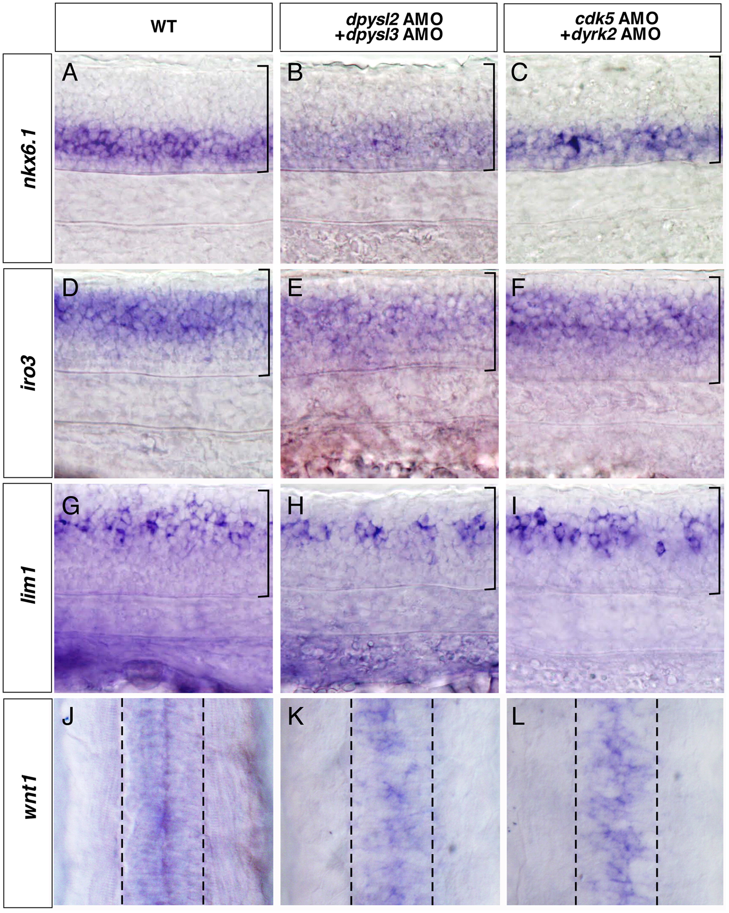
Fig. 5 Loss of function of cdk5, dyrk2, dpysl2, and dpysl3 specifically affected the patterning of the dorsal-most part of the spinal cord. (A–L) Expression of nkx6.1 (A–C), iro3 (D–F), lim1 (G–I), and wnt1 (J–L) in the wild-type embryos (A, D, G, J), the dpysl2 and dpysl3 double morphants (B, E, H, K), and the cdk5 and dyrk2 double morphants (C, F, I, L) at 28 hpf, anterior left (A–I), anterior top (J–L). In the wild-type embryos, nkx6.1, iro3, and lim1 were expressed in the ventral, middle to dorsal, and dorsal part of the spinal cord, respectively (A, D, G). In the dorsal-most part of the spinal cord, wnt1 was expressed around the midline (J). In the dpysl2 and dpysl3 double morphants or the cdk5 and dyrk2 double morphants, the expression patterns of nkx6.1, iro3, and lim1 were similar to those of the wild-type embryos (B, C, E, F, H, I). However, the expression domain of wnt1 was abnormally distributed around the midline (K, L). Brackets in A–I and broken lines in J–L indicate the spinal cord.
Reprinted from Developmental Biology, 370(2), Tanaka, H., Morimura, R., and Ohshima, T., Dpysl2 (CRMP2) and Dpysl3 (CRMP4) phosphorylation by Cdk5 and DYRK2 is required for proper positioning of Rohon-Beard neurons and neural crest cells during neurulation in zebrafish, 223-236, Copyright (2012) with permission from Elsevier. Full text @ Dev. Biol.