Fig. 8
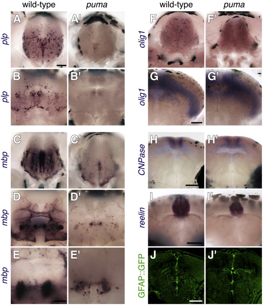
Fig. 8 Defects in oligodendrocyte number and patterning during the larval-to-adult transformation of puma mutants. (A, A′, B, B′) Wild-type zebrafish larvae exhibit many more plp+ oligodendrocytes in the brain than do puma mutants both during the middle larval period (A, A&prime& ~ 6.5 SSL) and the late larval period (B, B&prime& ~ 8.0 SSL). (C, C′, D, D′, E, E′) mbp expression also differs between wild-type and puma mutants during middle and later larval development (C, C′ and D, D′, respectively); a higher magnification view of different larvae is shown in E, E′. Note especially the absence of most myelinated fibers in the puma mutant and the concentration of mbp mRNA in cell bodies. (F, F′, G, G′) The early oligodendrocyte marker olig1 does not differ in expression between genotypes at middle or later larval stages, nor do several additional markers of particular cell lineages or activities (H–J; see text for details). Scale bars: in A, 100 μm, for A, A′, B, B′, C, C′, D, D′, F, F′. In E, 40 μm for E, E&prime& in G, 100 μm for G, G′ in H, 100 μm for H, H&prime& in J, 100 μm for I, J&prime& in J, 100 μm for J, K′.
Reprinted from Developmental Biology, 346(2), Larson, T.A., Gordon, T.N., Lau, H.E., and Parichy, D.M., Defective adult oligodendrocyte and Schwann cell development, pigment pattern, and craniofacial morphology in puma mutant zebrafish having an alpha tubulin mutation, 296-309, Copyright (2010) with permission from Elsevier. Full text @ Dev. Biol.

