
Fig. S3
- ID
- ZDB-IMAGE-081202-30
- Genes
- Publication
- Ma et al., 2007 - Jagged2a-notch signaling mediates cell fate choice in the zebrafish pronephric duct
- All Figures
- Figures for Ma et al., 2007
|
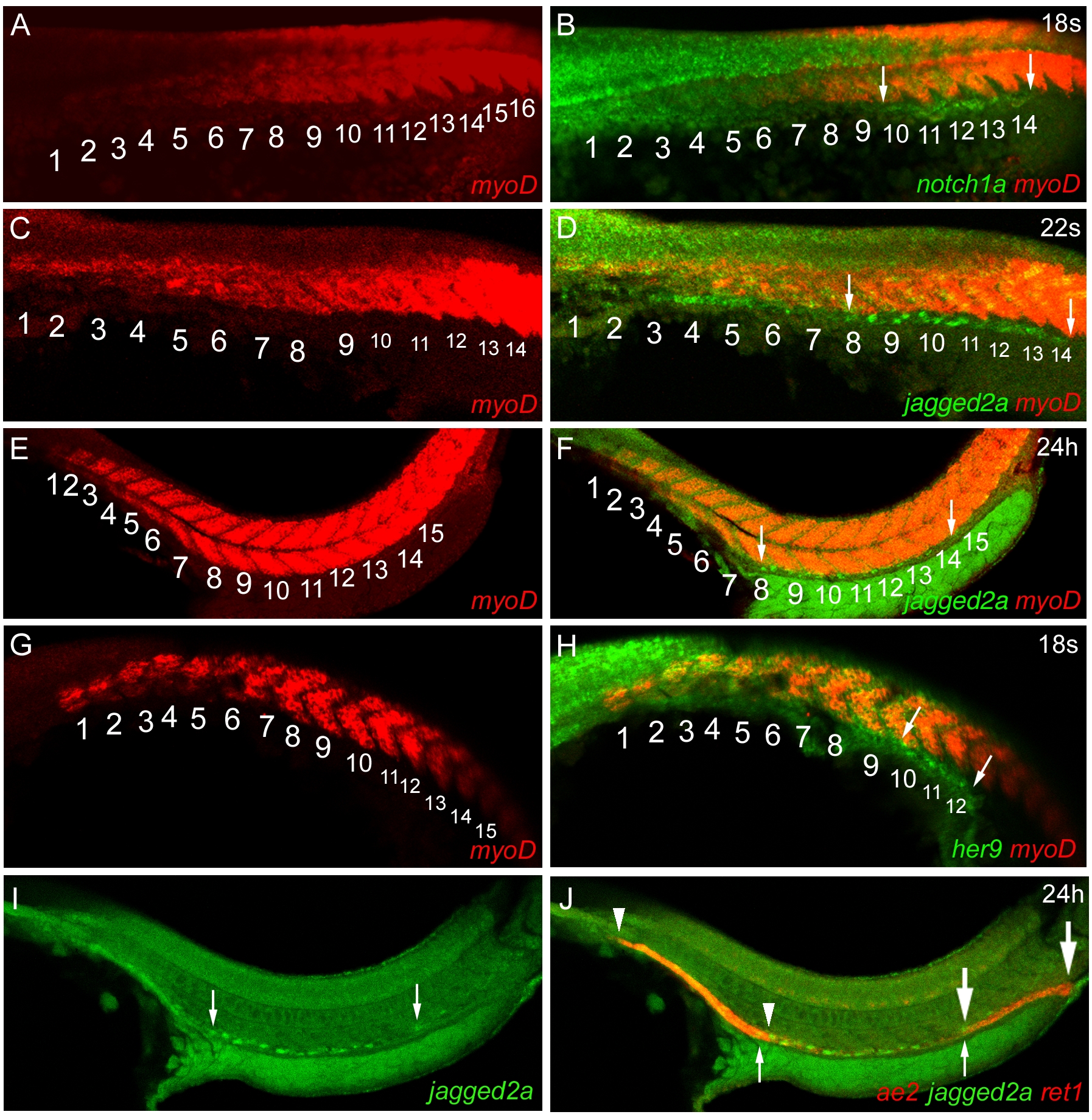
Fig. S3 notch1a, jagged2a, and her9 Are Expressed in the Distal Duct at the Time of Cell-Fate Determination
(A and B) Fluorescent double in situ hybridization of notch1a and myoD [93] revealed that notch1a is expressed in the pronephric duct spanning from somite 10 to 14 (arrows) at 18 ss.
(C and D) Fluorescent double in situ hybridization of jagged2a and myoD revealed that mosaic jagged2a expression is found in the pronephric duct spanning from somite 8 to 14 (arrows) at 22 ss.
(E and F) Fluorescent double in situ hybridization of jagged2a and myoD revealed that jagged2a-expressing single cells are found in the pronephric duct spanning from somite 8 to 14 (arrows) at 24 hpf.
(G and H) Fluorescent double in situ hybridization of her9 and myoD revealed that her9 is expressed in the pronephric duct spanning from somite 10 to 12 (arrows) at 18 ss.
(I and J) Fluorescent double in situ hybridization of jagged2a (green), slc4a2/ae2 (red, anterior), and ret1 (red, posterior) revealed that jagged2a-expressing single cells are found in the distal duct between the proximal duct (marked by slc4a2/ae2; [27]) and the cloaca (marked by ret1; [11]). Small arrows demarcate the jagged2a-expressing single cell domain, arrowheads demarcate the slc4a2/ae2-expressing domain, and big arrows demarcate the ret1-expressing domain.