Fig. 1
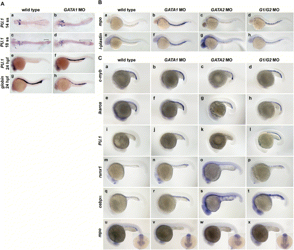
Fig. 1 Loss of Gata1 Results in Expanded Myelopoiesis(A) pu.1 expression in control and gata1 MO-injected embryos at 14 somites (a, b), 18 somites (c, d), and 24 hpf (e, f). Embryos (a–d) were flat-mounted with anterior (left) and posterior (right). gata1 MO-injected embryos express pu.1 at 18 somites (d, brackets) and 24 hpf (f) while wild-type embryos have downregulated pu.1 transcripts (c, brackets, e) in the ICM precursors. pu.1 is also expressed in anterior myeloid precursors (a–f). gata1 MO-injected embryos have reduced expression of βe1 globin (h) in the ICM at 24 hpf compared to wild-types (g).(B) The number of cells expressing mpo and l-plastin are increased in the vessels of gata1 morphants (b, f, d, h) compared to wild-type embryos (a, e) and gata2 morphants (c, g). g1/g2 morphants (d, h) have decreased numbers of mpo- and l-plastin-expressing cells compared to the gata1 morphants alone (b, f).(C) Expression of c-myb (a–d) and ikaros (e–h) is maintained in the ICM cells of gata1, gata2, and g1/g2 morphants at 20 somites. pu.1 expression persists in the ICM of gata1 (j) and g1/g2 (l) morphants, but not gata2 morphants (k) or wild-type embryos (i). Expression of runx1 (m–p) and cebpα (q–t) persists in gata1 (n, r) and g1/g2 morphants (p, t) but not wild-type embryos (m, q) or gata2 morphants (o, s) at 22 hpf. cebpα is expressed at low levels in some ICM cells and in gut endoderm (q–t). ICM cells expressing mpo at 22 hpf are reduced in gata2 morphants (w) and lost in gata1 (v) and g1/g2 morphants (x) compared to wild-type embryos (u). Insets (u–x) are of cranial views of the same embryos showing normal mpo-expressing cells in wild-type (u) and gata1 morphants (v) and reduced numbers of mpo-expressing cells in gata2 (w) and g1/g2 morphants (x).
Reprinted from Developmental Cell, 8(1), Galloway, J.L., Wingert, R.A., Thisse, C., Thisse, B., and Zon, L.I., Loss of gata1 but not gata2 converts erythropoiesis to myelopoiesis in zebrafish embryos, 109-116, Copyright (2005) with permission from Elsevier. Full text @ Dev. Cell

