
Fig. 1
- ID
- ZDB-IMAGE-060808-502
- Genes
- Publication
- Stigloher et al., 2006 - Segregation of telencephalic and eye-field identities inside the zebrafish forebrain territory is controlled by Rx3
- All Figures
- Figures for Stigloher et al., 2006
|
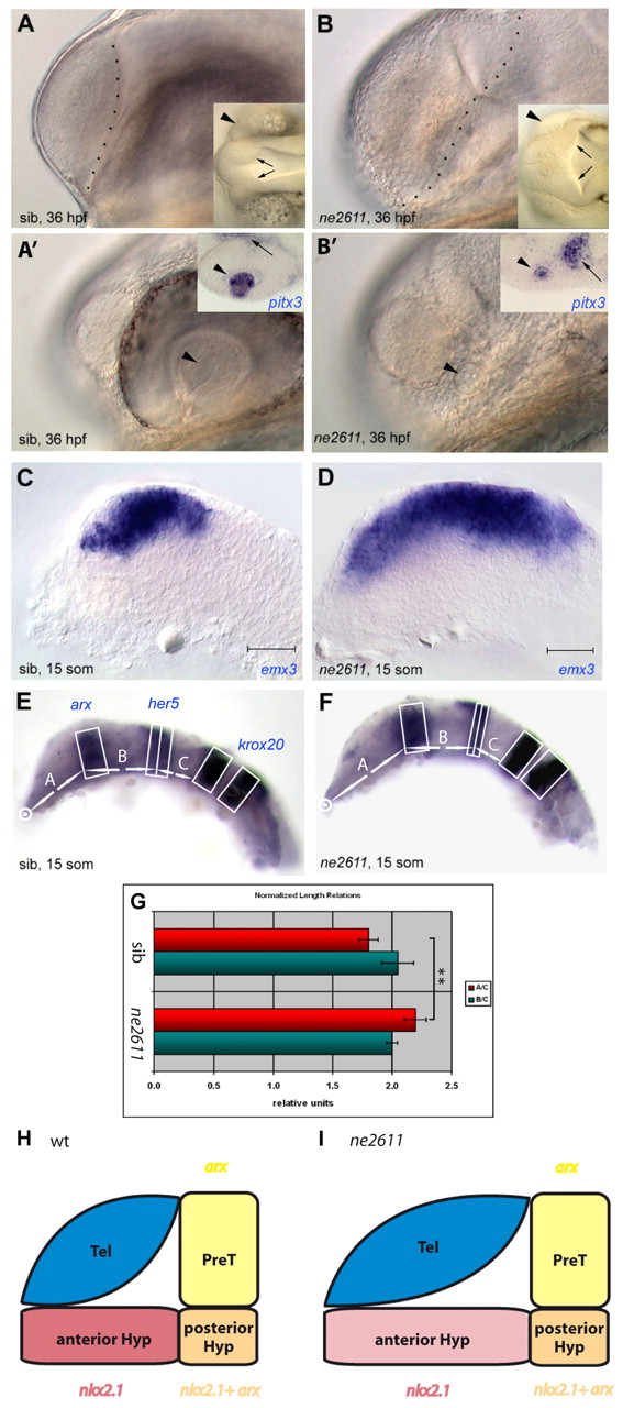
Fig. 1 Enlarged telencephalon and lack of retina in ne2611. All views are lateral, anterior left. (A-B') Compared morphology of ne2611 (B,B') and wild-type siblings (sib; A,A') at 36 hpf; views with parasagittal and lateral focus, respectively. (A,B) Note the enlarged telencephalon, delimited by a dashed line, in ne2611. Insets are dorsal views of the same embryos showing absence of the retina (arrowheads) and an expanded diencephalic ventricle (arrows) in ne2611. (A',B') Note the maintenance of a lens (arrowhead) in ne2611, albeit smaller than in wild-type siblings. Insets show expression of pitx3, molecularly identifying the lens (arrowheads) in both genotypes. The adjacent domain of pitx3 expression (small arrows) is hypothalamic, it is displaced towards the lens in ne2611 due to the absence of eyes. (C,D) Compared expression of the telencephalic marker emx3 at the 15-somite stage in ne2611 (D) and wild-type siblings (C); scale bar: 0.02 mm. The mutants display a massive anteroposterior enlargement of the emx3 domain. (E-G) Relative anteroposterior sizes of the anteriormost forebrain (domain A) versus prethalamus, thalamus, pretectum and midbrain (domain B) at 15 somites in ne2611 (E) compared with wild-type siblings (F). (G) Measurements are normalized to the size of the anterior hindbrain (domain C), and the domains are defined with the genes indicated in E. Bars indicate s.e.m. The difference in A/C length between wild type and ne2611 is statistically significant (two-sample independent Student's t-test, P values are given in the text). (H,I) Schematic representation of the size of the different anterior forebrain territories in wild type versus ne2611 siblings at 15 somites. The genes used as landmarks are color coded (yellow: arx only, prethalamus; red: nkx2.1 only, anterior hypothalamus; orange: arx+nkx2.1, posterior hypothalamus). Prethalamus and posterior hypothalamus are unchanged in the mutants. The anterior hypothalamus appears elongated, but might be simply stretched (hence the lighter red color) by the anteroposterior enlargement of the telencephalon. Later, the anterior hypothalamus appears reduced or missing (see also Fig. S1I,J in the supplementary material).