
Fig. 3
- ID
- ZDB-FIG-260805-107
- Publication
- Yi et al., 2026 - HUWE1 targets mitochondria via RMC1 to promote neurodevelopment
- Other Figures
- All Figure Page
- Back to All Figure Page
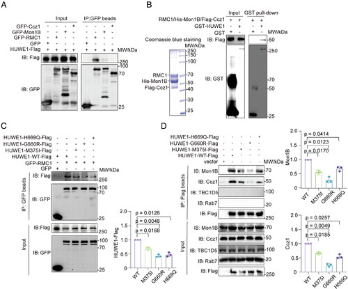
HUWE1 M375I, G660R, and H669Q variants impair HUWE1-RMC1 interaction. (A) GFP-tagged constructs encoding RMC1, Mon1B, Ccz1, and HUWE1-WT-Flag were transfected in HEK293T cells, GFP vector as the control. 24 h later, cells were collected and subjected to immunoprecipitation using GFP beads and immunoblotting. (B) GST and GST-HUWE1 were expressed and purified using HEK293T cells, then incubated with purified RMC1–Mon1–Ccz1 complex and subjected to GST pull-down. GST, GST-HUWE1, and Flag-Ccz1 were detected by antibodies against GST and Flag, respectively. (C) HEK293T cells were cotransfected with GFP-RMC1 and Flag-tagged HUWE1 WT, M375I, G660R, or H669Q, with empty GFP vector included as a control. At 24 h after transfection, cell lysates were prepared and GFP-associated protein complexes were isolated using GFP beads, followed by immunoblot analysis with the indicated antibodies. The relative amounts of bound HUWE1 were calculated as the ratio of coprecipitated HUWE1-Flag to GFP-RMC1 signal intensity. Quantification was performed with ImageJ and normalized to the WT group. Data are shown as mean ± SD from three independent experiments. Statistical significance was analyzed by one-way ANOVA followed by multiple-comparison analysis. (D) HEK293T cells were transfected with Flag-tagged HUWE1 WT, M375I, G660R, or H669Q, with empty Flag vector used as a control. At 24 h after transfection, HUWE1-Flag protein complexes were enriched using Flag beads and examined by immunoblotting. The relative amounts of bound Mon1B and Ccz1 were quantified by normalizing their signal intensities to the corresponding HUWE1-Flag signals and are plotted relative to WT. Data represent mean ± SD from n = 3 independent experiments, P values were calculated using one-way ANOVA followed by multiple-comparison analysis. |