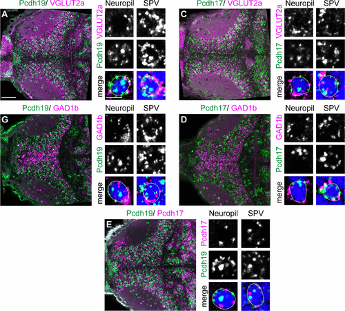
Expression of pcdh19 and pcdh17 in the optic tectum.
A HCR-ISH showing the expression of pcdh19 (green) and vglut2a (magenta), which is a marker of excitatory neurons (left panel, scale bar = 50 μm.). Pcdh19 is expressed in excitatory neurons both in neuropil interneurons (middle column, scale bar = 5 μm), and neurons in the stratum periventriculare (SPV) neurons (right column). DAPI is shown in blue. The overlap of green spots and blue shows up is displayed as cyan. White line highlights an individual cell. B HCR-ISH showing the expression of pcdh19 (green) and gad1b (magenta), which is a marker of inhibitory neurons (left panel). Pcdh19 is expressed in inhibitory neurons both in neuropil interneurons (middle column), and neurons in the stratum periventriculare (SPV) neurons (right column). C HCR-ISH showing the expression of pcdh17 (green) and vglut2a (magenta), which is a marker of excitatory neurons (left panel). Pcdh17 is expressed in excitatory neurons both in neuropil interneurons (middle column), and neurons in the stratum periventriculare (SPV) neurons (right column). D HCR-ISH showing the expression of pcdh17 (green) and gad1b (magenta), which is a marker of inhibitory neurons (left panel). Pcdh17 is expressed in inhibitory neurons both in neuropil interneurons (middle column), and neurons in the stratum periventriculare (SPV) neurons (right column). E HCR-ISH showing the expression of pcdh19 (green) and pcdh17 (magenta) in the otpic tectum (left panel). Pcdh19 and Pcdh17 are expressed in distinct but overlapping sets of neurons. Examples of co-expression are shown both for neuropil interneurons (middle column), and neurons in the stratum periventriculare (SPV) neurons (right column).
|
