
Figure 4
- ID
- ZDB-FIG-260508-20
- Publication
- Pannese et al., 2026 - Impaired collagen deposition in the absence of ERp44
- Other Figures
- All Figure Page
- Back to All Figure Page
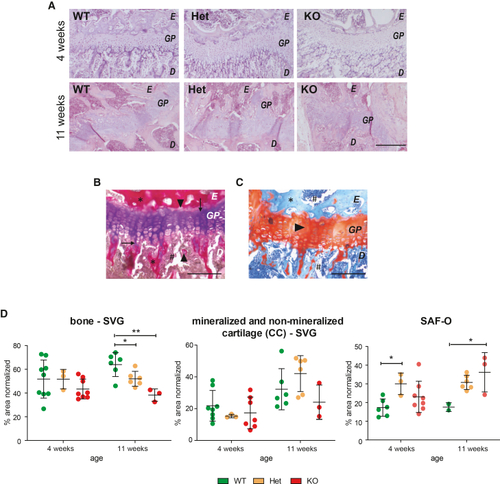
Delayed bone mineralization in ERp44 KO mice by means of histological staining of the femoral growth plate (A) Hematoxylin and eosin staining of the growth plate at 4 and 11 weeks of age (top and bottom, respectively). Growth plates (GP) are larger in both Het and KO with respect to WT control mice at 4 weeks of age. E, epiphysis; D, diaphysis. Scale bars, 200 μm. (B) Example of growth plate in Stevenel’s/Van Gieson (SVG) staining at higher magnification (bone in red color: asterisks; mineralized cartilage in pink color: arrowheads; cartilage in violet color: arrows; bone marrow in brown color: hashtag) to show details of epiphyseal growth plate components. (C) Example of growth plate in Safranin-O/light green section at higher magnification (bone in light blue color: asterisk; cartilage in red color: arrowhead; bone marrow in dark blue color: hashtag). E, epiphysis; GP, growth plate; D, diaphysis. Scale bars, 25 μm. (D) SVG quantifications at 4 weeks of age ( |