
Figure 6
- ID
- ZDB-FIG-260508-22
- Publication
- Pannese et al., 2026 - Impaired collagen deposition in the absence of ERp44
- Other Figures
- All Figure Page
- Back to All Figure Page
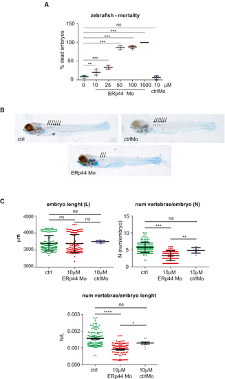
ERp44 morpholinos induced delayed spine mineralization in zebrafish embryos (A) ERp44 MO injection in fertilized zebrafish eggs induces a dose-dependent embryo mortality. Five independent experiments (13 embryos/condition in each experiment). Mortality in embryos injected with control morpholinos (blue color) is equivalent to that of uninjected morpholinos (green color), near to 0% in both groups, contrary to MO group (red color). Average ± SD, One-way ANOVA with Dunnett’s multiple comparison test (∗∗ (B) Images of zebrafish embryos, at 5 days after fertilization, untreated (ctrl), treated with 10 μM control MO (ctrlMO) or with 10 μM MO anti ERp44 (ERp44MO). Staining was performed with Alcian blue (light blue, cartilage) and Alizarin red (violet, mineralized bone). Black arrows point to mineralized vertebrae (in violet). ERp44 MO treatment decreases the number of mineralized vertebrae in developing embryos. (C) Graphs report embryo length, number of mineralized vertebrae, and the ratio “number of vertebrae/embryo length” for the three groups (control in green, Morpholinos treatment in red, control MO in blue). Each dot represents one embryo, from independent experiments. Average ± SD, Student’s |
| Fish: | |
|---|---|
| Knockdown Reagent: | |
| Observed In: | |
| Stage: | Day 5 |