
Figure 4
- ID
- ZDB-IMAGE-260508-23
- Publication
- Pannese et al., 2026 - Impaired collagen deposition in the absence of ERp44
- All Figures
- Figures for Pannese et al., 2026
|
Figure 4
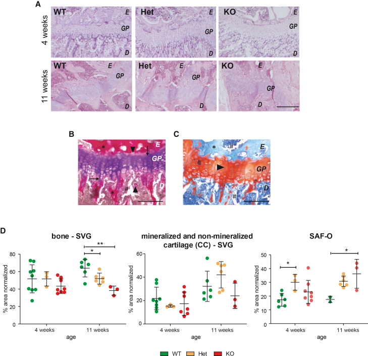
Delayed bone mineralization in ERp44 KO mice by means of histological staining of the femoral growth plate
(A) Hematoxylin and eosin staining of the growth plate at 4 and 11 weeks of age (top and bottom, respectively). Growth plates (GP) are larger in both Het and KO with respect to WT control mice at 4 weeks of age. E, epiphysis; D, diaphysis. Scale bars, 200 μm.
(B) Example of growth plate in Stevenel’s/Van Gieson (SVG) staining at higher magnification (bone in red color: asterisks; mineralized cartilage in pink color: arrowheads; cartilage in violet color: arrows; bone marrow in brown color: hashtag) to show details of epiphyseal growth plate components.
(C) Example of growth plate in Safranin-O/light green section at higher magnification (bone in light blue color: asterisk; cartilage in red color: arrowhead; bone marrow in dark blue color: hashtag). E, epiphysis; GP, growth plate; D, diaphysis. Scale bars, 25 μm.
(D) SVG quantifications at 4 weeks of age (