
Figure 7
- ID
- ZDB-FIG-260501-115
- Publication
- De Rycke et al., 2026 - Systematic Disruption of Zebrafish Fibrillin Genes Identifies a Translational Zebrafish Model for Marfan Syndrome
- Other Figures
- All Figure Page
- Back to All Figure Page
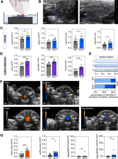
Cardiac Abnormalities of Adult fbn3–/– ZF (A) Schematic representation of the two-dimensional transthoracic echocardiography of adult ZF. The ZF is anesthetized, positioned with its ventral side upward, and submerged in water. The echocardiography measurements are made with an ultrasound probe, in abdominocranial axis (ACX) view for color flow Doppler (CFD) analysis (red) and in longitudinal view (LCX) for ventricle and BA measurements (dark red). (B) Tracings of the posterior walls of the ventricle (blue) and BA (purple) of WT ZF (left) and fbn3–/– ZF (right) (6 and 8 mpf). (C) Dimensions of the ventricle during diastole (area;d, volume;d) and systole (area;s, volume;s) (n = 12-14). (D) Measurements of BA volume, area, and diameter while in maximal relaxation (at the time of bulboventricular valve contraction) (n = 11-13). (E) Representative 20-second cardiac rhythm tracings with the associated qualitative score (top). Distribution of cardiac rhythm scores for different genotypes (n = 12) (bottom). (F) CFD recordings of inflow (orange) and outflow (blue) of blood into/from the ventricle in WT ZF (top) and fbn3–/– ZF (bottom), with an example of regurgitant blood flow seen in some mutants (n = 2 of 14). (G) Quantification of inflow (orange) and outflow (blue) areas and regurgitation fractions from the CFD data (n = 10-14). Values are mean ± SEM. Statistical test analysis: unpaired Student’s t-test (C, D, and G, inflow area), Mann-Whitney U test (G, outflow area, regurgitation fraction IN, regurgitation fraction OUT), and the Fisher exact test E. ∗P < 0.05, ∗∗P < 0.01, ∗∗∗P < 0.001. ns = nonsignificant; other abbreviations as in |
| Fish: | |
|---|---|
| Observed In: | |
| Stage: | Adult |