
FIGURE
Fig. 2
- ID
- ZDB-FIG-260409-22
- Publication
- Tasnim et al., 2025 - Functional divergence of Tbx2a and Tbx2b in zebrafish heart development
- Other Figures
- All Figure Page
- Back to All Figure Page
Fig. 2
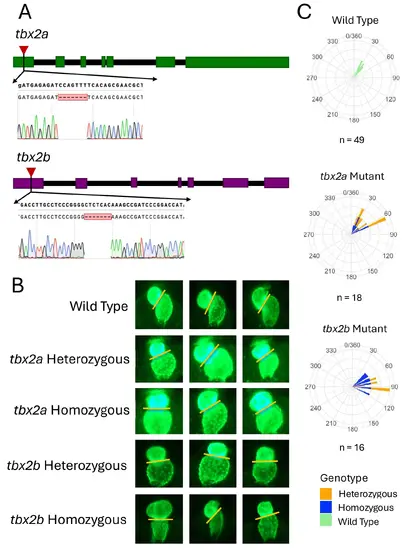
Heart looping defects in tbx2a and tbx2b mutant zebrafish 48 hpf. A Schematic of CRISPR-Cas9 mutations of tbx2a and tbx2b. Each colored block indicates an exon for that respective gene. B Fluorescent imaging of GFP-labeled hearts from myl7: GFP transgenic embryos at 48 hpf. C Polar histograms displaying the distribution of heart looping angles in wild-type (WT), tbx2a mutant, and tbx2b mutant zebrafish at 48 h post-fertilization (hpf). Each bar represents the frequency of observed looping angles, color-coded by genotype: Wild Type (light green), Heterozygous (orange), and Homozygous (blue) |
Expression Data
Expression Detail
Antibody Labeling
Phenotype Data
| Fish: | |
|---|---|
| Observed In: | |
| Stage: | Long-pec |
Phenotype Detail
Acknowledgments
This image is the copyrighted work of the attributed author or publisher, and
ZFIN has permission only to display this image to its users.
Additional permissions should be obtained from the applicable author or publisher of the image.
Full text @ EvoDevo