|
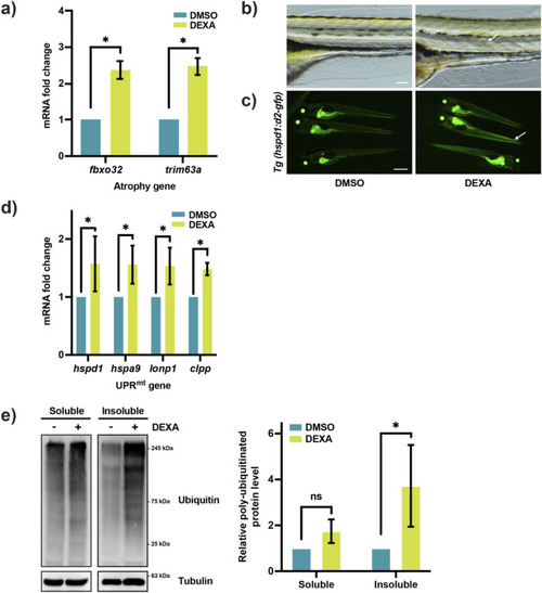
UPRmt is activated during muscle atrophy in zebrafish larvae. (a) qRT-PCR analysis of dexamethasone (DEXA)-treated zebrafish larvae, showing the expression of the atrophy genes fbxo32 and trim63a (*p < 0.05, t-test, two-tailed, error bar indicates s.d.). (b) Bright-field images of myotome from DEXA-treated zebrafish larvae at 6 dpf. Arrow indicates disorganized muscle fibers. Scale bar represents 0.1 mm. (c) Fluorescence images of the UPRmt reporter zebrafish treated with DEXA. Arrows indicate elevated fluorescent signals in the skeletal muscle. Scale bar represents 1 mm. (d) qRT-PCR analysis of DEXA-treated zebrafish larvae showing the expression of the UPRmt genes hspd1, hspa9, lonp1, and clpp (*p < 0.05, t-test, two-tailed, error bar indicates s.d.). (e) Western blot and quantification of poly-ubiquitinated proteins in the detergent-soluble and -insoluble fractions of the protein lysates from DEXA-treated zebrafish larvae (*p < 0.05, t-test, two-tailed, error bar indicates s.d.).
|