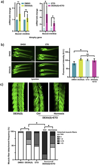
Inhibition of the mFAO sensitizes muscle to atrophic stress. (a) qRT-PCR analysis of zebrafish larvae treated with DEXA(S) and/or etomoxir (ETO) showing the expression of the atrophy genes fbxo32 and trim63a (DEXA(S) indicates a short-course DEXA treatment. *p < 0.05, t-test, two-tailed, error bar indicates s.d.). (b) Representative fluorescent images of LysoView-stained zebrafish larvae treated with DEXA(S) and/or ETO. Histogram showing quantification of fluorescent intensity (white rectangle indicates the area chosen for the fluorescence quantification. Scale bar represents 1 mm. *p < 0.05, one-way ANOVA and Tukey’s multiple comparison test; error bar indicates s.d.). (c) Representative confocal microscopy images of Alexa488-phalloidin-stained zebrafish larvae treated with DEXA(S) and/or ETO with or without the hormesis treatment (arrows indicate examples of detached muscle fibers). Boxplot shows quantification of larvae with 0, 1–2, 3–15, or >15 detached muscle fibers (*p < 0.05, Chi-square test. Scale bar represents 50 µM).
|
