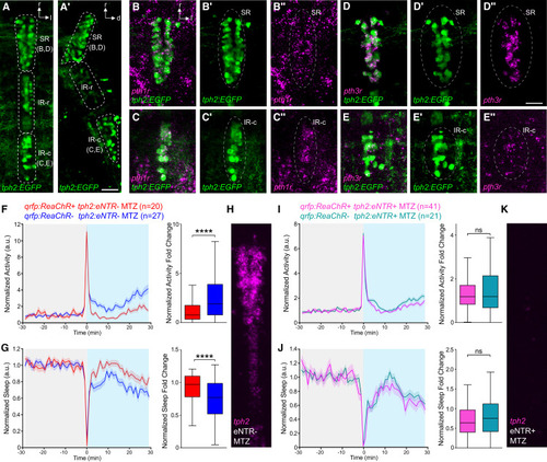
Sleep induced by optogenetic stimulation of Pth4 neurons is abolished by ablation of the 5HT raphe (A and A′) 5HT raphe neurons labeled in Tg(tph2:EGFP) fish include the SR, IR-r, and IR-c subregions, as seen in dorsal (A) and lateral (A′) views. (B–E″) HCR shows that pth1r (B–C″) and pth3r (D–E″) are both expressed in the SR (pth1r: 97/226 SR neurons express pth1r in 5 fish; pth3r: 312/422 SR neurons express pth3r in 11 fish). Both receptors are also expressed in the IR-c (pth1r: 76/90 IR-c neurons express pth1r in 5 fish; pth3r: 59/82 IR-c neurons show faint expression of pth3r in 4 fish). Raphe neurons are indicated with dashed ovals. Scale bars, 25 μm. (F–K) Optogenetic stimulation of Pth4 neurons in Tg(qrfp:ReaChR) fish suppresses locomotor activity and increases sleep in MTZ-treated eNTR-negative fish (F and G) with intact raphe (H), but not in MTZ-treated Tg(tph2:eNTR) fish (I and J) with ablated raphe (K), compared with ReaChR-negative fish. Mean ± SEM line plots and Tukey boxplots are shown. n = number of fish. ∗∗∗∗p < 0.0001, ns, p > 0.05 by Mann-Whitney U test. r, rostral; l, lateral; d, dorsal. See also Figures S2 and S5.
|
