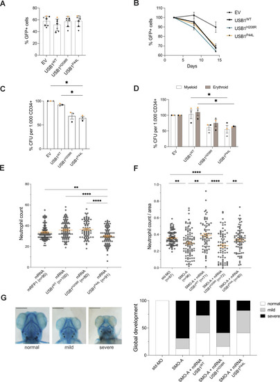
The USB1P44Lvariant impacts myeloid differentiation in vitro and in vivo. (A) Percentage of GFP+ cells in human CD34+ cells at day 2.5 following the transduction step (n = 7). Bars and error bars are the averages of the percentage of alive GFP+ cells and SD from seven independent experiments. (B) Percentage of GFP+ cells at days 8 and 14 normalized to the GFP+ population at day 2.5. Error bars are SEM from a minimum of two independent experiments (n = 2). (C) Colony-forming unit (CFU) potential of myeloid differentiation (n = 3). (D) CFU potential of myeloid (white bars) and erythroid (brown bars) differentiation (n = 3). (D) were evaluated after an 8-day culture. Bars and error bars are the averages of the percentage of the indicated populations and SEM from three independent experiments. Ordinary one-way ANOVA statistical analysis was performed. *P < 0.05. Statistically nonsignificant differences were not annotated. (E) Neutrophil count at 2 dpf in zebrafish overexpressing indicated USB1 variant RNAs (n = 4 biological replicates, in orange mean ± SEM). Ordinary one-way ANOVA statistical analysis was performed. **P < 0.01 and ****P < 0.0001. Statistically nonsignificant differences were not annotated. (F) SMO-A morphants injected with indicated USB1 variants (n = 4 biological replicates, in orange mean ± SEM). Ordinary one-way ANOVA statistical analysis was performed on log2-transformed data. *P < 0.05, **P < 0.01, ***P < 0.001, and ****P < 0.0001. Statistically nonsignificant differences were not annotated. (G) Classification of Alcian blue staining highlights morphological alterations at 5 dpf (n = 3). From left to right: ventral view of a control embryo (left) and representative pictures of the mild (middle) and severe (right). Scale bar = 250 μm.
|
