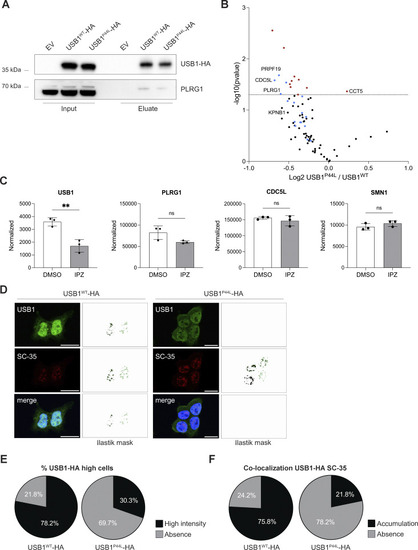
USB1 de novo variant impacts USB1 protein interactome. (A) HEK293T-USB1-HA lysates were immunoprecipitated using magnetic bead-bound anti-HA antibodies and analyzed by immunoblotting to validate the co-immunoprecipitation mass spectrometry results (n = 4). EV, empty vector. (B) Volcano plot of co-immunoprecipitation mass spectrometry experiments performed with HEK293T cells overexpressing USB1-HA variants (n = 4). Red dots denote statistically significantly enriched proteins. Known USB1 interactors are marked in blue. (C) Mass spectrometry analysis of nuclear fraction of DMSO- and IPZ-treated NB-4 cells (n = 3) (17). Proteomics data were obtained at PRIDE (PRoteomics IDEntification Database) under accession number PXD056172. An unpaired t test was performed for USB1 and its interactors (PLRG1, CDC5L, and SMN1). ns, nonsignificant differences (P ≥ 0.05), **P < 0.01. (D) Representative confocal microscopy images for HEK293T cells stably expressing USB1WT or USB1P44L variant (n = 3). SC-35 was included to visualize the nuclear speckles. Scale bar = 15 µm. For each image, pixel classification was performed using a machine learning Ilastik model, and the resulting mask is displayed. For the USB1-HA signal, we trained the model to create a mask of high-intensity signal voxels. (E) Percentage of cells presenting USB1-HA high-intensity signal in the nuclei (n = 3). (F) Signal overlap between USB1-HA and SC-35 was quantified using 3D pixel classification via an Ilastik machine learning model, focusing on high-intensity USB1-HA pixels. A threshold of 27 voxels was applied to define a cellular compartment. Semiautomated quantification was executed in Fiji (RRID:SCR_002285). IPZ, importazole. Source data are available for this figure: SourceData F4.
|
