FIGURE

Figure 6.

ID
ZDB-FIG-260105-108
Publication
Michno et al., 2025 - Pneumolysin-dependent and independent non-canonical autophagy processes mediate host defense against pneumococcal infection
Other Figures
All Figure Page
Back to All Figure Page
Figure 6.

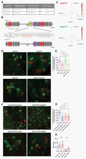
Tecpr1a, a likely ortholog of human TECPR1, controls ply-mediated autophagic response to pneumococci. (A) Table summarizing the alignment of zebrafish Tecpr1a and Tecpr1b proteins to the human TECPR1 protein. Columns display percentage positives (identical amino acids, as well as conserved or semiconserved substitutions) and length of the proteins. Alignments sequence similarities are shown for the full-length protein and the AIR domain, which exhibits the greatest divergence between the zebrafish paralogues (Tecpr1a and Tecpr1b). (B) Schematic representation of zebrafish Tecpr1a and Tecpr1b proteins, showing their domains: DysF – Dysferlin, AIR – ATG5-interacting region, and PH – Pleckstrin homology domain. Alignment of the AIR domain amino acid sequence of human TECPR1 and the zebfafish orthologs, Tecpr1a and Tecpr1b, is shown between the schematics. Symbols denote: (*) identical residues, (:) conserved substitutions, and (.) semiconserved substitutions. (C) Gene expression of adult zebrafish leukocytes determined using the zebrafish blood atlas [48]. Each dot represents a separate scRnaseq sample (cell); replicates were performed across multiple zebrafish wild-type and transgenic strains. Each arm of the schematic represents a separate blood cell population (labeled). Deeper color indicates higher expression (log10 scale bars described for each gene). (D) Representative confocal images of control, tecpr1a, tecpr1b and tecpr1a tecpr1b double knockdown CMV:GFP-Lc3 larvae infected systemically with approximately 1600 CFU of mKate2-labeled D39 δcps S. pneumoniae fixed at 2 hpi. (E) Quantification of Lc3 associations with intracellular S. pneumoniae within infected phagocytes of fixed tecpr1a, tecpr1b and tecpr1a tecpr1b double knockdown CMV:GFP-Lc3 larvae. Data are shown as mean ± standard deviation (SD). n ≥ 17 zebrafish larvae were analyzed. (F) representative confocal images of control or tecpr1a knockdown CMV:GFP-Lc3 larvae infected systemically with approximately 1600 CFU of mKate2-labeled ply-positive (D39 Δcps) or ply-negative (D39 Δcps Δply) S. pneumoniae fixed at 2 hpi. (G) Quantification of Lc3 associations with intracellular ply-positive or ply-negative S. pneumoniae within infected phagocytes of control or tecpr1a knockdown CMV:GFP-Lc3 larvae fixed at 2 hpi. Data are shown as mean ± standard deviation (SD). Data obtained from two independent experiments. (H) Numbers of viable colony forming units (CFUs) over time of control and tecpr1a knockdown larvae. Data obtained from two independent experiments.

Expression Data

Expression Detail
Antibody Labeling
Phenotype Data

Phenotype Detail
Acknowledgments
This image is the copyrighted work of the attributed author or publisher, and ZFIN has permission only to display this image to its users. Additional permissions should be obtained from the applicable author or publisher of the image. Full text @ Autophagy