|
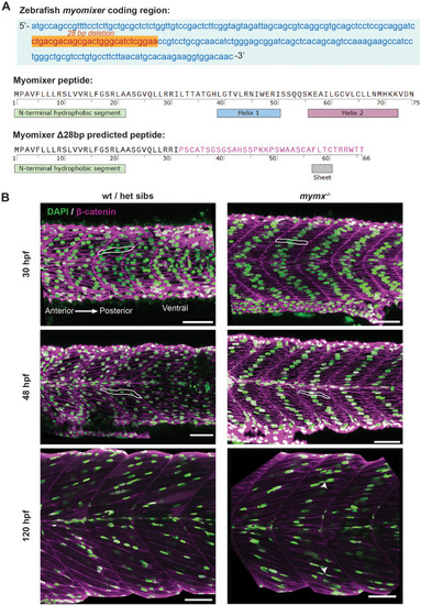
Loss of mymx function disrupted embryonic and larval skeletal muscle fibre morphology. (A) Top: nucleotide sequence of the mymx coding region in zebrafish showing the 28 bp deletion (orange) by CRISPR-Cas9 gene editing. Bottom: amino acid sequences and predicted secondary structural elements of native and mutant Mymx peptides, with the altered amino acids of the extracellular domain shown in pink. (B) Wild-type siblings (left column) and mymx−/− (right column) embryos at 30, 48 and 120 hpf, immunostained for nuclei (DAPI, green) and cell membranes (β-catenin, magenta) to visualise the skeletal muscle fibres in the developing myotomes. Scale bars: 50 µm. Example cells highlighted with white outlines. Fused cells in mymx−/− embryos at 120 hpf highlighted by white arrowheads.
|