FIGURE

Fig. 9

ID
ZDB-FIG-250906-54
Publication
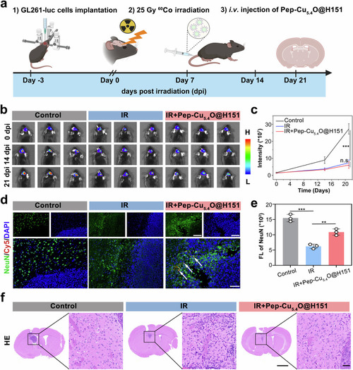
Shang et al., 2025 - Understanding the toxicity induced by radiation-triggered neuroinflammation and the on-demand design of targeted peptide nanodrugs
Other Figures
All Figure Page
Back to All Figure Page
Fig. 9

Neuroprotection by the Pep-Cu5.4O@H151 nanoplatform at the tumor–normal tissue interface during radiotherapy for glioblastoma. a Schematic illustration of the experimental design: The GL261 orthotopic glioma model was established via stereotactic inoculation, followed by fractionated radiotherapy (5 Gy × 5 days) and concurrent nanodrug administration (5 mg/kg, i.v., 14 days). b Representative in vivo bio-fluorescence images of tumor volume at 0, 14 and 21 days post-treatment. c Quantitative analysis of tumor bio-fluorescence intensity (n = 3 mice; one-way ANOVA). d Immunofluorescence staining of NeuN⁺ neurons. NeuN⁺ neurons (green); Pep-Cu5.4O@H151 nanodrugs (red, white arrows indicate neuron-specific accumulation); DAPI (blue). e Quantitative analysis of NeuN⁺ neuron fluorescence intensity (n = 3 fields; one-way ANOVA). f HE staining of mouse brains. The left panel shows the whole-brain image, and the right panel presents a magnified view of the area within the black box in the left panel. All the statistical data are presented as the means ± SEMs. Scale bars: 20 μm (d), 1000 μm (f, right), and 20 μm (f, left)

Expression Data

Expression Detail
Antibody Labeling
Phenotype Data

Phenotype Detail
Acknowledgments
This image is the copyrighted work of the attributed author or publisher, and ZFIN has permission only to display this image to its users. Additional permissions should be obtained from the applicable author or publisher of the image. Full text @ Signal Transduct Target Ther