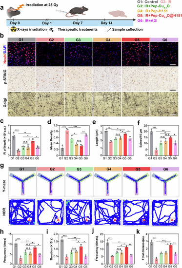
In vivo therapeutic effects of Pep-Cu5.4O@H151 nanodrugs on RIBI. a Schematic illustration of the in vivo experimental design. The mice were exposed to fractionated radiation (5 Gy × 5 times, cumulative 25 Gy) to induce RIBI. Starting 24 h post-IR, the mice were administered 150 μL of the test formulations via tail vein injection daily for 14 consecutive days. The experimental groups included the following: (1) control (PBS injection, G1); (2) IR group (radiation, G2); (3) Pep-Cu5.4O group (radiation + 5 mg/kg Pep-Cu5.4O, G3); (4) Pep-H151 group (radiation + 5 mg/kg Pep-H151, G4); (5) Pep-Cu5.4O@H151 group (radiation + 5 mg/kg total concentration, with 2.5 mg/kg Pep, 2.5 mg/kg Cu5.4O, and 1.25 mg/kg H151, G5); and (6) ADI group (radiation + 10 mg/kg idebenone as a positive control, G6). Each group consisted of six mice. b Representative NeuN immunofluorescence staining images (Alexa Fluor 594, red) and representative STING immunohistochemistry (DAB) images of the cortex. Images of Golgi-Cox-stained pyramidal neurons. c Quantification of NeuN⁺ fluorescence intensity normalized to that of DAPI⁺-stained nuclei (n = 5 fields; one-way ANOVA). d Quantification of the mean STING⁺ density (n = 3 fields; one-way ANOVA). e, f Dendritic complexity analysis via the Sholl assay: radial length (e) and branch density (f) (n = 3 fields; one-way ANOVA). g Representative heatmaps of Y-maze and novel object recognition tests in 16 wpi mice. h Frequency of entries into the novel arm (N) in the Y-maze test. i Duration of time spent in the novel arm (N) of the Y-maze. j Frequency of interactions with the novel object (Object A) during the novel object recognition test. k Total movement distance across the testing area. All the statistical data are presented as the means ± SEMs. Scale bar: 20 μm (b)
|
